 Explore
 Validate
 Learn
Learn Western blot
 Immunohistochemistry
Antibody data
- Antibody Data
- Antigen structure
- References [1]
- Comments [0]
- Validations
- Immunohistochemistry [1]
Submit
Validation data
Reference
Comment
Report error
- Product number
- MAB3264 - Provider product page

- Provider
- Novus Biologicals
- Product name
- Rat Monoclonal Cadherin-13 Antibody
- Antibody type
- Monoclonal
- Description
- Protein A or G purified from hybridoma culture supernatant. Detects human Cadherin-13 in direct ELISAs and Western blots. In direct ELISAs and Western blots, no cross-reactivity with recombinant human (rh) E-Cadherin, rhN-Cadherin, rhP-Cadherin, rhVE-Cadherin, rhCadherin-8, -11, -12 or -17 is observed.
- Reactivity
- Human, Mouse
- Host
- Rat
- Conjugate
- Unconjugated
- Isotype
- IgG
- Vial size
- 100 ug
- Concentration
- LYOPH
- Storage
- Use a manual defrost freezer and avoid repeated freeze-thaw cycles. 12 months from date of receipt, -20 to -70 degreesC as supplied. 1 month, 2 to 8 degreesC under sterile conditions after reconstitution. 6 months, -20 to -70 degreesC under sterile conditions after reconstitution.
Submitted references Positive feedback regulation between adiponectin and T-cadherin impacts adiponectin levels in tissue and plasma of male mice.
Matsuda K, Fujishima Y, Maeda N, Mori T, Hirata A, Sekimoto R, Tsushima Y, Masuda S, Yamaoka M, Inoue K, Nishizawa H, Kita S, Ranscht B, Funahashi T, Shimomura I
Endocrinology 2015 Mar;156(3):934-46
Endocrinology 2015 Mar;156(3):934-46
No comments: Submit comment
Supportive validation
- Submitted by
- Novus Biologicals (provider)
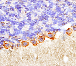
- Main image
- Experimental details
- Cadherin-13 in Mouse Brain. Cadherin-13 was detected in immersion fixed frozen sections of mouse brain (cerebellum) using Rat Anti-Human Cadherin-13 Monoclonal Antibody (Catalog # MAB3264) at 25 µg/mL overnight at 4 °C. Tissue was stained using the Anti-Rat HRP-DAB Cell & Tissue Staining Kit (brown; Catalog # CTS017) and counterstained with hematoxylin (blue). Specific staining was localized to Purkinje cells. View our protocol for Chromogenic IHC Staining of Frozen Tissue Sections.
