 Explore
 Validate
 Learn
Learn Western blot
Antibody data
- Antibody Data
- Antigen structure
- References [0]
- Comments [0]
- Validations
- Western blot [2]
- Immunocytochemistry [6]
- Immunohistochemistry [4]
- Flow cytometry [1]
Submit
Validation data
Reference
Comment
Report error
- Product number
- MA5-24853 - Provider product page

- Provider
- Invitrogen Antibodies
- Product name
- PHD1 Recombinant Rabbit Monoclonal Antibody (3G4)
- Antibody type
- Monoclonal
- Antigen
- Recombinant full-length protein
- Reactivity
- Human, Mouse, Rat
- Host
- Rabbit
- Isotype
- IgG
- Antibody clone number
- 3G4
- Vial size
- 100 µL
- Concentration
- 1 mg/mL
- Storage
- Store at 4°C short term. For long term storage, store at -20°C, avoiding freeze/thaw cycles.
No comments: Submit comment
Supportive validation
- Submitted by
- Invitrogen Antibodies (provider)
- Main image

- Experimental details
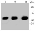
- Western blot analysis of PHD1 in Lane 1: HeLa whole cell lysate, Lane 2: PC12 whole cell lysate, Lane 3: NIH/3T3 whole cell lysate. Samples were incubated with PHD1 monoclonal antibody (Product # MA5-24853) at a dilution of 1:1,000.
- Submitted by
- Invitrogen Antibodies (provider)
- Main image
- Experimental details
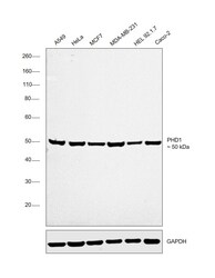
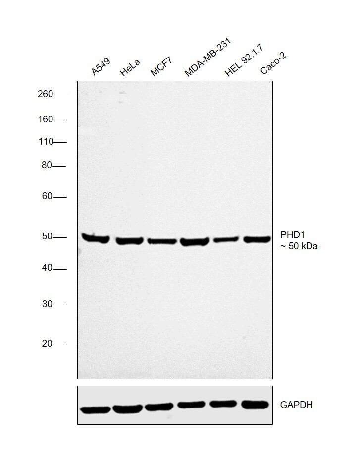
- Western blot was performed using Anti-PHD1 Recombinant Rabbit Monoclonal Antibody (Product # MA5-24853) and a 50 kDa band corresponding to PHD1 was observed across all the cell lines tested. Modified whole cell extracts (1% SDS) (30 µg lysate) of A549 (Lane 1), HeLa (Lane 2), MCF7 (Lane 3), MDA-MB-231 (Lane 4), HEL 92.1.7 (Lane 5) and Caco-2 (Lane 6) were electrophoresed using Novex® NuPAGE® 4-12% Bis-Tris Protein Gel (Product # NP0322BOX). Resolved proteins were then transferred onto a nitrocellulose membrane (Product # IB23001) by iBlot® 2 Dry Blotting System (Product # IB21001). The blot was probed with the primary antibody (1:1000 dilution) and detected by chemiluminescence with Goat anti-Rabbit IgG (H+L), Superclonal™ Recombinant Secondary Antibody, HRP (Product # A27036, 1:4000 dilution) using the iBright FL 1000 (Product # A32752). Chemiluminescent detection was performed using Novex® ECL Chemiluminescent Substrate Reagent Kit (Product # WP20005).
Supportive validation
- Submitted by
- Invitrogen Antibodies (provider)
- Main image
- Experimental details
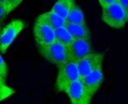
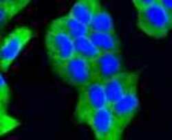
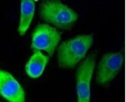
- Immunofluorescence analysis of PHD1 in HeLa cells (green). Samples were treated with paraformaldehyde, permeabilized with 0.25% Triton and PBS, and incubated with PHD1 monoclonal antibody (Product # MA5-24853).

- Submitted by
- Invitrogen Antibodies (provider)
- Main image
- Experimental details
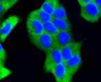
- Immunofluorescence analysis of PHD1 in HeLa cells (green). Samples were treated with paraformaldehyde, permeabilized with 0.25% Triton and PBS, and incubated with PHD1 monoclonal antibody (Product # MA5-24853).
- Submitted by
- Invitrogen Antibodies (provider)
- Main image
- Experimental details
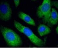
- Immunofluorescence analysis of PHD1 in SKOV-3 cells (green). Samples were treated with paraformaldehyde, permeabilized with 0.25% Triton and PBS, and incubated with PHD1 monoclonal antibody (Product # MA5-24853).
- Submitted by
- Invitrogen Antibodies (provider)
- Main image

- Experimental details
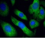
- Immunofluorescence analysis of PHD1 in HeLa cells (green). Samples were treated with paraformaldehyde, permeabilized with 0.25% Triton and PBS, and incubated with PHD1 monoclonal antibody (Product # MA5-24853).

- Submitted by
- Invitrogen Antibodies (provider)
- Main image
- Experimental details
- Immunofluorescence analysis of PHD1 in A549 cells (green). Samples were treated with paraformaldehyde, permeabilized with 0.25% Triton and PBS, and incubated with PHD1 monoclonal antibody (Product # MA5-24853).

- Submitted by
- Invitrogen Antibodies (provider)
- Main image
- Experimental details
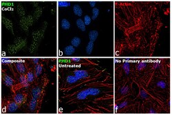
- Immunofluorescence analysis of PHD1 was performed using 70% confluent log phase HeLa cells treated with Cobalt chloride. The cells were fixed with 4% paraformaldehyde for 10 minutes, permeabilized with 0.1% Triton™ X-100 for 15 minutes, and blocked with 2% BSA for 1 hour at room temperature. The cells were labeled with PHD1 Recombinant Rabbit Monoclonal Antibody (3G4) (Product # MA5-24853) at 1:100 dilution in 0.1% BSA, incubated at 4 degree Celsius overnight and then labeled with Goat anti-Rabbit IgG (H+L) Superclonal™ Recombinant Secondary Antibody, Alexa Fluor® 488 conjugate (Product # A27034) at a dilution of 1:2000 for 45 minutes at room temperature (Panel a: green). Nuclei (Panel b: blue) were stained with ProLong™ Diamond Antifade Mountant with DAPI (Product # P36962). F-actin (Panel c: red) was stained with Rhodamine Phalloidin (Product # R415). Panel d represents the merged image showing increase in Nuclear localization. Panel e represents untreated cells with Nuclear localization. Panel f represents control cells with no primary antibody to assess background. The images were captured at 60X magnification.
Supportive validation
- Submitted by
- Invitrogen Antibodies (provider)
- Main image

- Experimental details
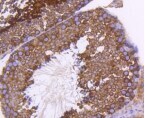
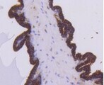
- Immunohistochemistry analysis of PHD1 in paraffin-embedded mouse testis tissue. Samples were incubated with PHD1 monoclonal antibody (Product # MA5-24853) followed by hematoxylin.
- Submitted by
- Invitrogen Antibodies (provider)
- Main image

- Experimental details
- Immunohistochemistry analysis of PHD1 in paraffin-embedded mouse prostate tissue. Samples were incubated with PHD1 monoclonal antibody (Product # MA5-24853) followed by hematoxylin.
- Submitted by
- Invitrogen Antibodies (provider)
- Main image

- Experimental details
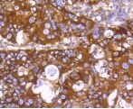
- Immunohistochemistry analysis of PHD1 in paraffin-embedded human lung cancer tissue. Samples were incubated with PHD1 monoclonal antibody (Product # MA5-24853) followed by hematoxylin.
- Submitted by
- Invitrogen Antibodies (provider)
- Main image

- Experimental details
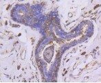
- Immunohistochemistry analysis of PHD1 in paraffin-embedded human breast carcinoma tissue. Samples were incubated with PHD1 monoclonal antibody (Product # MA5-24853) followed by hematoxylin.
Supportive validation
- Submitted by
- Invitrogen Antibodies (provider)
- Main image

- Experimental details
- Flow cytometry analysis of PHD1 in HeLa cells (blue) and compared with an unlabeled control (cells without incubation with primary antibody; red). Samples were incubated with PHD1 monoclonal antibody (Product # MA5-24853) at a dilution of 1:50 followed by Alexa Fluor 488-conjugated goat anti rabbit IgG.
