 Explore
 Validate
 Learn
Learn Western blot
 Immunocytochemistry
 Immunohistochemistry
Antibody data
- Antibody Data
- Antigen structure
- References [0]
- Comments [0]
- Validations
- Western blot [3]
- ELISA [3]
- Immunohistochemistry [8]
Submit
Validation data
Reference
Comment
Report error
- Product number
- LS-C745284 - Provider product page

- Provider
- LSBio
- Product name
- APP / Beta Amyloid Precursor Antibody LS-C745284
- Antibody type
- Polyclonal
- Description
- Affinity purified
- Reactivity
- Human, Mouse
- Host
- Rabbit
- Isotype
- IgG
- Storage
- Store vial at -20°C or below prior to opening. Dilute 1:10 to minimize loss. Store the vial at -20°C or below after dilution. Avoid freeze-thaw cycles.
No comments: Submit comment
Supportive validation
- Submitted by
- LSBio (provider)
- Enhanced method
- Genetic validation
- Main image

- Experimental details
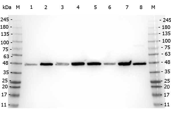
- Western Blot of rabbit anti-Beta Amyloid antibody. Marker: Opal Pre-stained ladder Lane 1: HEK293 lysate Lane 2: HeLa Lysate Lane 3: MCF-7 Lysate Lane 4: Jurkat Lysate Lane 5: A431 Lysate Lane 6: LNCaP Lysate Lane 7: A-172 Lysate Lane 8: NIH/3T3 Lysate Load: 35 µg per lane. Primary antibody: Beta Amyloid antibody at 1:5,000 for overnight at 4°C. Secondary antibody: Peroxidase rabbit secondary antibody at 1:30,000 for 60 min at RT. Blocking Buffer: 1% Casein-TTBS for 30 min at RT. Predicted/Observed size: 40 kDa, 40 kDa for AHA1. Other band(s): N/A.
- Submitted by
- LSBio (provider)
- Enhanced method
- Genetic validation
- Main image
- Experimental details
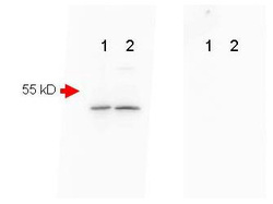
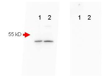
- Mouse Brain (Lane 1) and Mouse Spinal Chord (Lane 2) were run on a 4-20% gradient gel, Blocked in 1% BSA-TBS-T 30 min RT and probed with Rb-a-Beta Amyloid 1:1000 in 1% BSA-TBS-T o/n 4°C. HRP Gt-a-Rb 611-103-122 Lot#21231 1:40,000 in MB-070 30 min RT. FEMTOMAX chemiluminescent substrate was used for detection of a 40-50 kD band consistent with a higher MW precursor form of beta amyloid. A secondary Ab only control (Shown right) showed no detectable signal.
- Submitted by
- LSBio (provider)
- Enhanced method
- Genetic validation
- Main image

- Experimental details
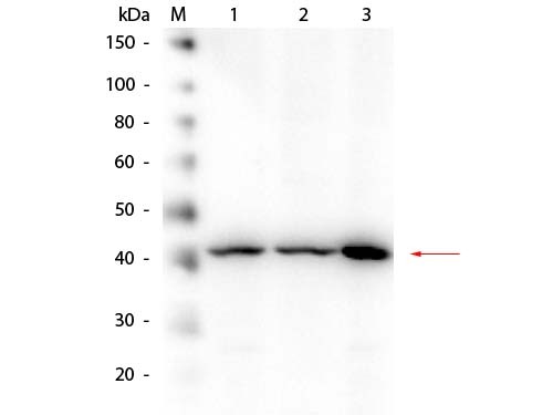
- Western Blot of rabbit anti-Beta Amyloid Antibody. Lane 1: HEK293 WCL. Lane 2: Mouse Brain WCL. Lane 3: A-172 WCL. Load: 10.0 µg per lane. Primary antibody: Beta Amyloid Antibody at 1:1,000 overnight at 4°C. Secondary antibody: Peroxidase Conjugated Goat-a-Rabbit IgG at 1:40,000 for 30 min at RT. Block: MB-070 for 30 min at RT. Predicted/Observed size: 40 kDa, 40 kDa for Beta Amyloid.
Supportive validation
- Submitted by
- LSBio (provider)
- Enhanced method
- Genetic validation
- Main image

- Experimental details
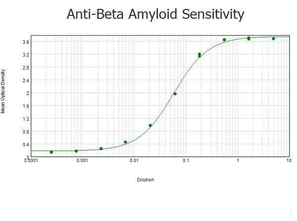
- ELISA results of purified Rabbit anti-Beta Amyloid Antibody tested against BSA-conjugated peptide of immunizing peptide. Each well was coated in duplicate with 0.1µg of conjugate. The starting dilution of antibody was 5µg/ml and the X-axis represents the Log10 of a 3-fold dilution. This titration is a 4-parameter curve fit where the IC50 is defined as the titer of the antibody. Assay performed using 3% fish gel, Goat anti-Rabbit IgG Antibody Peroxidase Conjugated (Min X Bv Ch Gt GP Ham Hs Hu Ms Rt & Sh Serum Proteins)
- Submitted by
- LSBio (provider)
- Main image
- Experimental details
- ELISA results of purified Rabbit anti-Beta Amyloid Antibody tested against BSA-conjugated peptide of immunizing peptide. Each well was coated in duplicate with 0.1µg of conjugate. The starting dilution of antibody was 5µg/ml and the X-axis represents the Log10 of a 3-fold dilution. This titration is a 4-parameter curve fit where the IC50 is defined as the titer of the antibody. Assay performed using 3% fish gel, Goat anti-Rabbit IgG Antibody Peroxidase Conjugated (Min X Bv Ch Gt GP Ham Hs Hu Ms Rt & Sh Serum Proteins)
- Submitted by
- LSBio (provider)
- Main image

- Experimental details
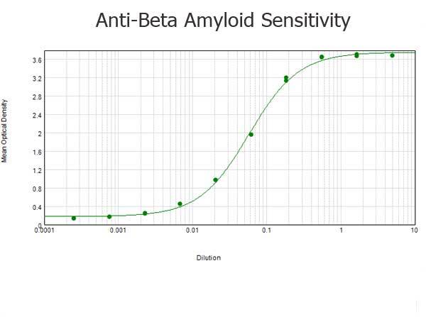
- ELISA results of purified Rabbit anti-Beta Amyloid Antibody tested against BSA-conjugated peptide of immunizing peptide. Each well was coated in duplicate with 0.1µg of conjugate. The starting dilution of antibody was 5µg/ml and the X-axis represents the Log10 of a 3-fold dilution. This titration is a 4-parameter curve fit where the IC50 is defined as the titer of the antibody. Assay performed using 3% fish gel, Goat anti-Rabbit IgG Antibody Peroxidase Conjugated (Min X Bv Ch Gt GP Ham Hs Hu Ms Rt & Sh Serum Proteins)

Enhanced validation
- Submitted by
- LSBio (provider)
- Enhanced method
- Genetic validation
- Main image
- Experimental details
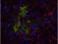
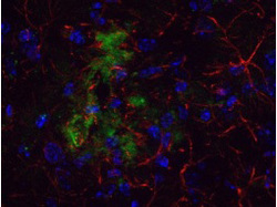
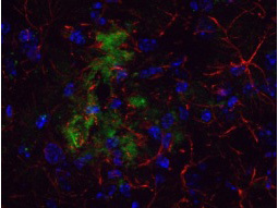
- Immunohistochemistical detection of beta Amyloid using Anti-Beta Amyloid Antibody on TG APP23 mouse brain cortex frozen sections. Anti-Beta Amyloid Antibody used at 1/200 and incubated for 2 hours in TBS/BSA/Tween/azide. Fluorescent labeled anti rabbit IgG was then added.

- Submitted by
- LSBio (provider)
- Enhanced method
- Genetic validation
- Main image

- Experimental details
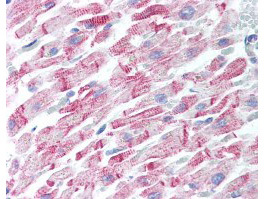
- Human Heart (formalin-fixed, paraffin-embedded) stained with Anti-Beta Amyloid Antibody at 5 ug/ml followed by biotinylated goat anti-rabbit IgG secondary antibody, alkaline phosphatase-streptavidin and chromogen.

- Submitted by
- LSBio (provider)
- Enhanced method
- Genetic validation
- Main image

- Experimental details
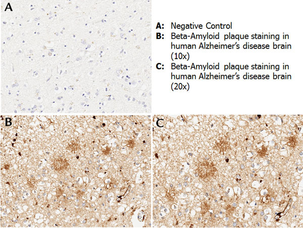
- Immunohistochemistry with anti-beta amyloid antibody showing amyloid beta plaque staining in human Alzheimer’s disease brain at 10x and 20x (B & C). Staining was performed on Leica Bond system using the standard protocol. Formalin fixed/paraffin embedded tissue sections were subjected to antigen retrieval with E1 (Leica Microsystems) retrieval solution for 20 min and then incubated with rabbit anti-beta amyloid antibody 600-401-253 at 1:100 dilution for 60 minutes. Biotinylated Anti-rabbit secondary antibody was used at 1:200 dilution to detect primary antibody. The reaction was developed using streptavidin-HRP conjugated compact polymer system and visualized with chromogen substrate, 3’3-diamino-benzidine substrate (DAB). The sections were then counterstained with hematoxylin to detect cell nuclei.
- Submitted by
- LSBio (provider)
- Enhanced method
- Genetic validation
- Main image

- Experimental details
- Immunohistochemistical detection of beta Amyloid using Anti-Beta Amyloid Antibody on TG APP23 mouse brain cortex frozen sections. Anti-Beta Amyloid Antibody used at 1/200 and incubated for 2 hours in TBS/BSA/Tween/azide. Fluorescent labeled anti rabbit IgG was then added.

- Submitted by
- LSBio (provider)
- Enhanced method
- Genetic validation
- Main image

- Experimental details
- Human Heart (formalin-fixed, paraffin-embedded) stained with Anti-Beta Amyloid Antibody at 5 ug/ml followed by biotinylated goat anti-rabbit IgG secondary antibody, alkaline phosphatase-streptavidin and chromogen.
- Submitted by
- LSBio (provider)
- Main image
- Experimental details
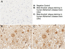
- Immunohistochemistry with anti-beta amyloid antibody showing amyloid beta plaque staining in human Alzheimer’s disease brain at 10x and 20x (B & C). Staining was performed on Leica Bond system using the standard protocol. Formalin fixed/paraffin embedded tissue sections were subjected to antigen retrieval with E1 (Leica Microsystems) retrieval solution for 20 min and then incubated with rabbit anti-beta amyloid antibody 600-401-253 at 1:100 dilution for 60 minutes. Biotinylated Anti-rabbit secondary antibody was used at 1:200 dilution to detect primary antibody. The reaction was developed using streptavidin-HRP conjugated compact polymer system and visualized with chromogen substrate, 3’3-diamino-benzidine substrate (DAB). The sections were then counterstained with hematoxylin to detect cell nuclei.

- Submitted by
- LSBio (provider)
- Main image
- Experimental details
- Immunohistochemistical detection of beta Amyloid using Anti-Beta Amyloid Antibody on TG APP23 mouse brain cortex frozen sections. Anti-Beta Amyloid Antibody used at 1/200 and incubated for 2 hours in TBS/BSA/Tween/azide. Fluorescent labeled anti rabbit IgG was then added.
- Submitted by
- LSBio (provider)
- Main image

- Experimental details
- Human Heart (formalin-fixed, paraffin-embedded) stained with Anti-Beta Amyloid Antibody at 5 ug/ml followed by biotinylated goat anti-rabbit IgG secondary antibody, alkaline phosphatase-streptavidin and chromogen.
