Explore
 Validate
 Learn
Learn Western blot
Antibody data
- Antibody Data
- Antigen structure
- References [1]
- Comments [0]
- Validations
- Western blot [1]
- Immunohistochemistry [7]
Submit
Validation data
Reference
Comment
Report error
- Product number
- HPA001462 - Provider product page

- Provider
- Atlas Antibodies
- Proper citation
- Atlas Antibodies Cat#HPA001462, RRID:AB_1078185
- Product name
- Anti-APP
- Antibody type
- Polyclonal
- Reactivity
- Human
- Host
- Rabbit
- Conjugate
- Unconjugated
- Antigen sequence
ANMISEPRISYGNDALMPSLTETKTTVELLPVNGE
FSLDDLQPWHSFGADSVPANTENEVEPVDARPAAD
RGLTTRPGSGLTNIKTEEISEVKMDAEFRHDSGYE
VHHQKLVFFAKDVGSN- Isotype
- IgG
- Vial size
- 100 µl
- Storage
- Store at +4°C for short term storage. Long time storage is recommended at -20°C.
Submitted references Candidate serological biomarkers for cancer identified from the secretomes of 23 cancer cell lines and the human protein atlas.
Wu CC, Hsu CW, Chen CD, Yu CJ, Chang KP, Tai DI, Liu HP, Su WH, Chang YS, Yu JS
Molecular & cellular proteomics : MCP 2010 Jun;9(6):1100-17
Molecular & cellular proteomics : MCP 2010 Jun;9(6):1100-17
No comments: Submit comment
Enhanced validation
- Submitted by
- Atlas Antibodies (provider)
- Enhanced method
- Recombinant expression validation
- Main image
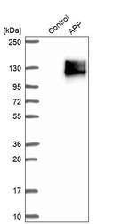
- Experimental details
- Western blot analysis in control (vector only transfected HEK293T lysate) and APP over-expression lysate (Co-expressed with a C-terminal myc-DDK tag (~3.1 kDa) in mammalian HEK293T cells, LY404408).
Enhanced validation
Supportive validation
- Submitted by
- Atlas Antibodies (provider)
- Enhanced method
- Orthogonal validation
- Main image
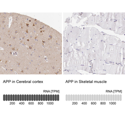
- Experimental details
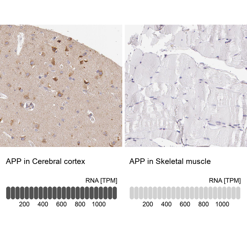
- Immunohistochemistry analysis in human cerebral cortex and skeletal muscle tissues using HPA001462 antibody. Corresponding APP RNA-seq data are presented for the same tissues.
- Sample type
- HUMAN
Supportive validation
- Submitted by
- Atlas Antibodies (provider)
- Main image
- Experimental details
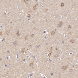
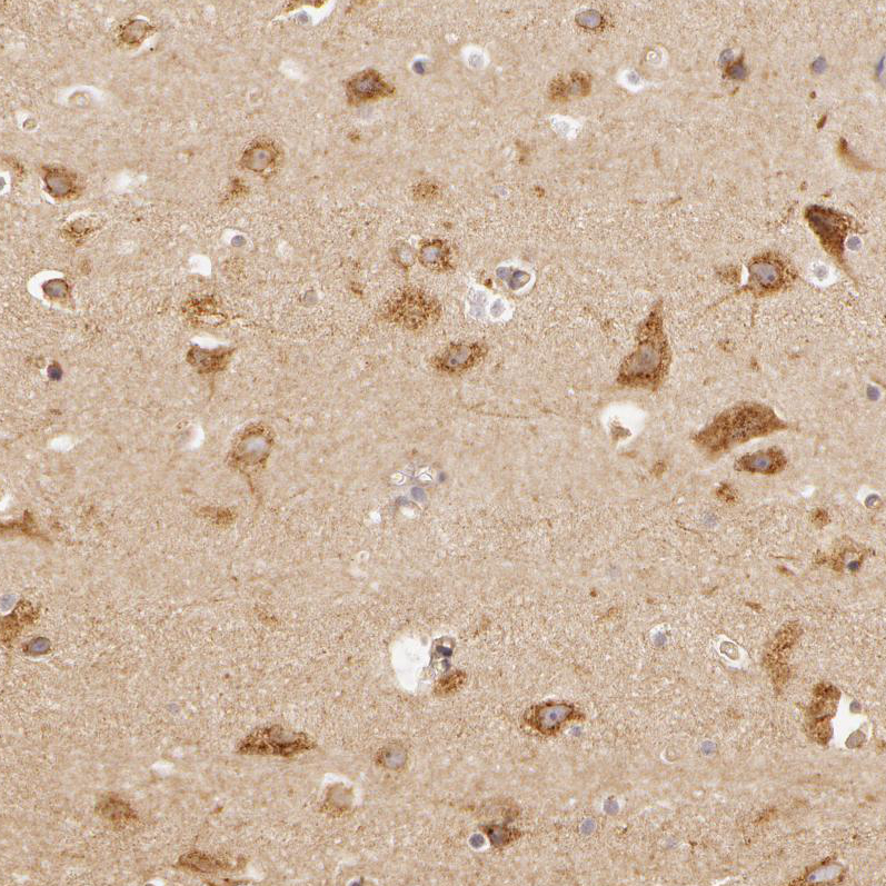
- Immunohistochemical staining of human cerebral cortex shows high expression.
- Sample type
- HUMAN
- Submitted by
- Atlas Antibodies (provider)
- Main image
- Experimental details
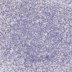
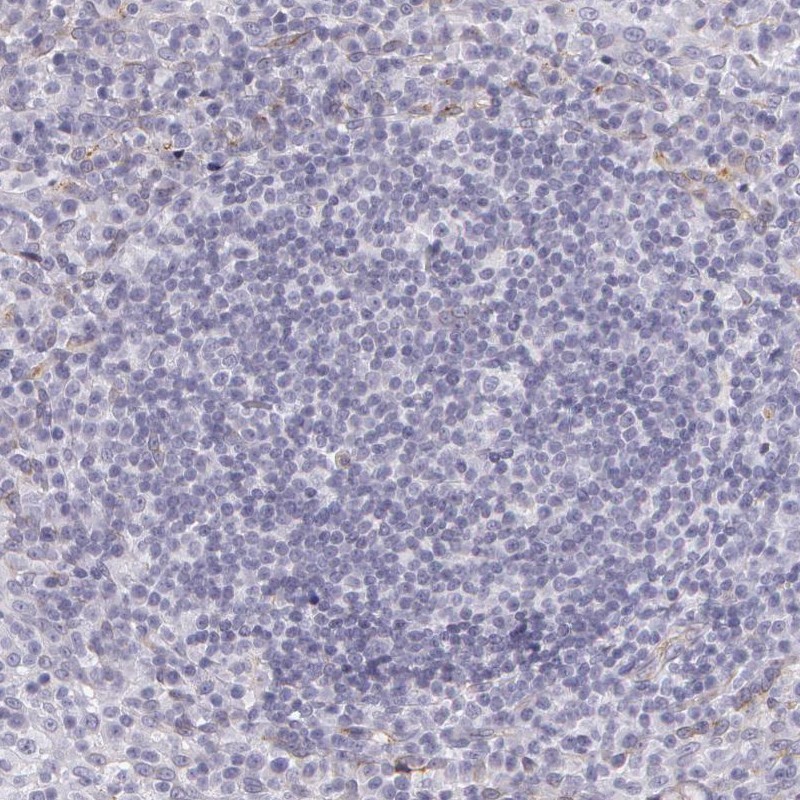
- Immunohistochemical staining of human tonsil shows low expression as expected.
- Sample type
- HUMAN
- Submitted by
- Atlas Antibodies (provider)
- Main image

- Experimental details
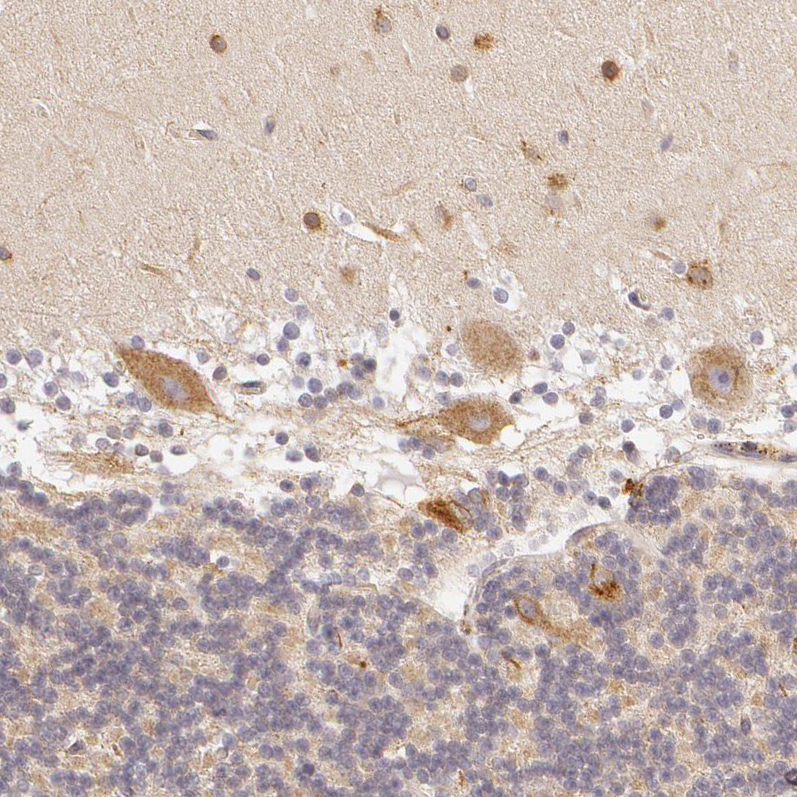
- Immunohistochemical staining of human hippocampus shows moderate granular positivity in cytoplasm in neurons.
- Sample type
- HUMAN
- Submitted by
- Atlas Antibodies (provider)
- Main image

- Experimental details
- Immunohistochemical staining of human cerebellum shows moderate granular positivity in cytoplasm in Purkinje cells.
- Sample type
- HUMAN
- Submitted by
- Atlas Antibodies (provider)
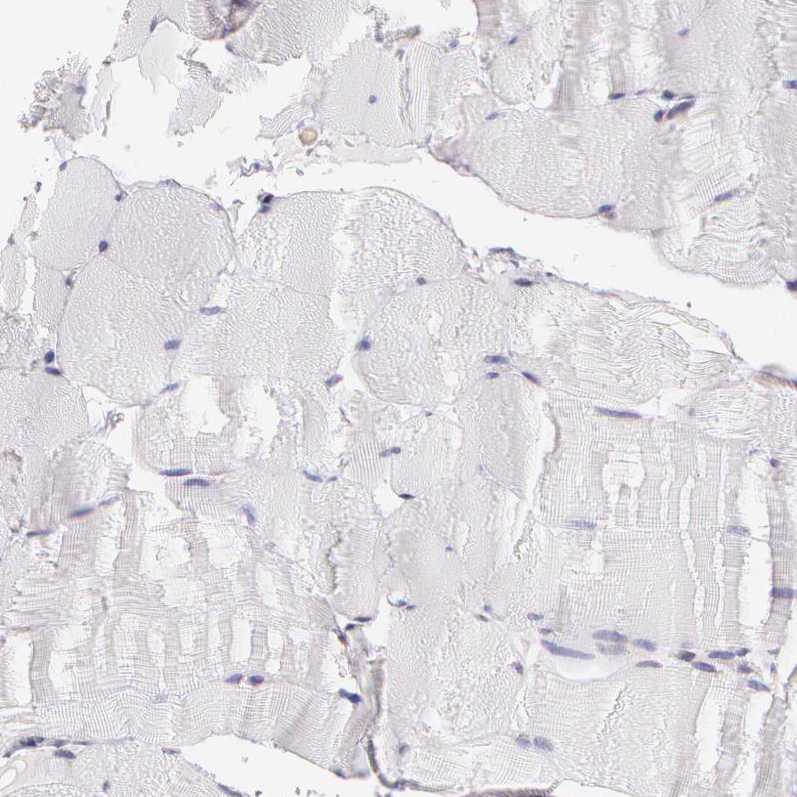
- Main image
- Experimental details
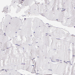
- Immunohistochemical staining of human skeletal muscle shows no positivity in myocytes as expected.
- Sample type
- HUMAN
- Submitted by
- Atlas Antibodies (provider)
- Main image

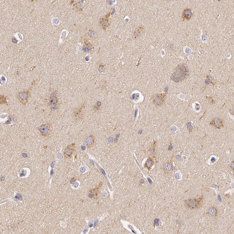
- Experimental details
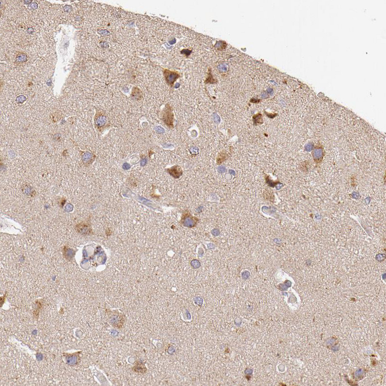
- Immunohistochemical staining of human cerebral cortex shows moderate granular positivity in cytoplasm in neurons.
- Sample type
- HUMAN