 Explore
 Validate
 Learn
Learn Western blot
Antibody data
- Antibody Data
- Antigen structure
- References [8]
- Comments [0]
- Validations
- Western blot [3]
- Immunohistochemistry [1]
Submit
Validation data
Reference
Comment
Report error
- Product number
- AF4439 - Provider product page

- Provider
- Novus Biologicals
- Product name
- Goat Polyclonal Contactin-2/TAG1 Antibody
- Antibody type
- Polyclonal
- Description
- Antigen Affinity-purified. Detects mouse and rat Contactin-2 in direct ELISAs. Detects human, mouse, and rat Contactin-2 in Western blots.
- Reactivity
- Human, Mouse, Rat
- Host
- Goat
- Conjugate
- Unconjugated
- Isotype
- IgG
- Vial size
- 100 ug
- Concentration
- LYOPH
- Storage
- Use a manual defrost freezer and avoid repeated freeze-thaw cycles. 12 months from date of receipt, -20 to -70 degreesC as supplied. 1 month, 2 to 8 degreesC under sterile conditions after reconstitution. 6 months, -20 to -70 degreesC under sterile conditions after reconstitution.
Submitted references Conditional ablation and conditional rescue models for Casq2 elucidate the role of development and of cell-type specific expression of Casq2 in the CPVT2 phenotype.
Phenotypically silent Cre recombination within the postnatal ventricular conduction system.
MUNC18-1 gene abnormalities are involved in neurodevelopmental disorders through defective cortical architecture during brain development.
Genome Stability by DNA Polymerase β in Neural Progenitors Contributes to Neuronal Differentiation in Cortical Development.
NOVA2-mediated RNA regulation is required for axonal pathfinding during development.
An aberrant sugar modification of BACE1 blocks its lysosomal targeting in Alzheimer's disease.
BACE1 activity regulates cell surface contactin-2 levels.
Secretome protein enrichment identifies physiological BACE1 protease substrates in neurons.
Flores DJ, Duong T, Brandenberger LO, Mitra A, Shirali A, Johnson JC, Springer D, Noguchi A, Yu ZX, Ebert SN, Ludwig A, Knollmann BC, Levin MD, Pfeifer K
Human molecular genetics 2018 May 1;27(9):1533-1544
Human molecular genetics 2018 May 1;27(9):1533-1544
Phenotypically silent Cre recombination within the postnatal ventricular conduction system.
Bhattacharyya S, Bhakta M, Munshi NV
PloS one 2017;12(3):e0174517
PloS one 2017;12(3):e0174517
MUNC18-1 gene abnormalities are involved in neurodevelopmental disorders through defective cortical architecture during brain development.
Hamada N, Iwamoto I, Tabata H, Nagata KI
Acta neuropathologica communications 2017 Nov 30;5(1):92
Acta neuropathologica communications 2017 Nov 30;5(1):92
Genome Stability by DNA Polymerase β in Neural Progenitors Contributes to Neuronal Differentiation in Cortical Development.
Onishi K, Uyeda A, Shida M, Hirayama T, Yagi T, Yamamoto N, Sugo N
The Journal of neuroscience : the official journal of the Society for Neuroscience 2017 Aug 30;37(35):8444-8458
The Journal of neuroscience : the official journal of the Society for Neuroscience 2017 Aug 30;37(35):8444-8458
NOVA2-mediated RNA regulation is required for axonal pathfinding during development.
Saito Y, Miranda-Rottmann S, Ruggiu M, Park CY, Fak JJ, Zhong R, Duncan JS, Fabella BA, Junge HJ, Chen Z, Araya R, Fritzsch B, Hudspeth AJ, Darnell RB
eLife 2016 May 25;5
eLife 2016 May 25;5
An aberrant sugar modification of BACE1 blocks its lysosomal targeting in Alzheimer's disease.
Kizuka Y, Kitazume S, Fujinawa R, Saito T, Iwata N, Saido TC, Nakano M, Yamaguchi Y, Hashimoto Y, Staufenbiel M, Hatsuta H, Murayama S, Manya H, Endo T, Taniguchi N
EMBO molecular medicine 2015 Feb;7(2):175-89
EMBO molecular medicine 2015 Feb;7(2):175-89
BACE1 activity regulates cell surface contactin-2 levels.
Gautam V, D'Avanzo C, Hebisch M, Kovacs DM, Kim DY
Molecular neurodegeneration 2014 Jan 9;9:4
Molecular neurodegeneration 2014 Jan 9;9:4
Secretome protein enrichment identifies physiological BACE1 protease substrates in neurons.
Kuhn PH, Koroniak K, Hogl S, Colombo A, Zeitschel U, Willem M, Volbracht C, Schepers U, Imhof A, Hoffmeister A, Haass C, Roßner S, Bräse S, Lichtenthaler SF
The EMBO journal 2012 Jun 22;31(14):3157-68
The EMBO journal 2012 Jun 22;31(14):3157-68
No comments: Submit comment
Supportive validation
- Submitted by
- Novus Biologicals (provider)
- Main image

- Experimental details
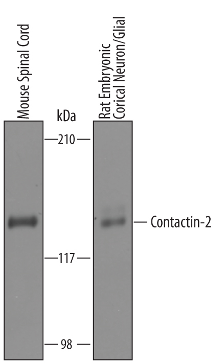
- Detection of Mouse and Rat Contactin-2/TAG1 by Western Blot. Western blot shows lysates of mouse spinal cord tissue and rat embryonic cortical neuron/glial cells. PVDF Membrane was probed with 1 µg/mL of Goat Anti-Mouse/Rat Contactin-2/TAG1 Polyclonal Antibody (Catalog # AF4439) followed by HRP-conjugated Anti-Goat IgG Secondary Antibody (Catalog # HAF019). A specific band was detected for Contactin-2/TAG1 at approximately 135 kDa (as indicated). This experiment was conducted under reducing conditions and using Immunoblot Buffer Group 8.
- Submitted by
- Novus Biologicals (provider)
- Main image

- Experimental details
- Detection of Mouse Contactin-2/TAG1 by Simple WesternTM. Simple Western lane view shows lysates of mouse spinal cord tissue, loaded at 0.2 mg/mL. A specific band was detected for Contactin-2/TAG1 at approximately 162 kDa (as indicated) using 10 µg/mL of Goat Anti-Mouse/Rat Contactin-2/TAG1 Antigen Affinity-purified Polyclonal Antibody (Catalog # AF4439) followed by 1:50 dilution of HRP-conjugated Anti-Goat IgG Secondary Antibody (Catalog # HAF109). This experiment was conducted under reducing conditions and using the 12-230 kDa separation system.

- Submitted by
- Novus Biologicals (provider)
- Main image

- Experimental details
- Detection of Human, Mouse, and Rat Contactin-2/TAG1 by Western Blot. Detection of Human, Mouse, and Rat Contactin-2/TAG1 by Western Blot.

Supportive validation
- Submitted by
- Novus Biologicals (provider)
- Main image

- Experimental details
- Contactin-2/TAG1 in Mouse Embryo. Contactin-2/TAG1 was detected in immersion fixed frozen sections of mouse embryo (E13) using Goat Anti-Mouse/Rat Contactin-2/TAG1 Antigen Affinity-purified Polyclonal Antibody (Catalog # AF4439) at 15 µg/mL overnight at 4 °C. Tissue was stained using the Anti-Goat HRP-DAB Cell & Tissue Staining Kit (brown; Catalog # CTS008) and counterstained with hematoxylin (blue). Specific staining was localized to muscle cells in proximity to ribs. View our protocol for Chromogenic IHC Staining of Frozen Tissue Sections.
