Explore
 Validate
 Learn
Learn Western blot
Antibody data
- Antibody Data
- Antigen structure
- References [1]
- Comments [0]
- Validations
- Western blot [2]
- Immunocytochemistry [1]
Submit
Validation data
Reference
Comment
Report error
- Product number
- AF7646 - Provider product page

- Provider
- R&D Systems
- Product name
- Human DNMT3B Antibody
- Antibody type
- Polyclonal
- Description
- Antigen Affinity-purified. Detects human DNMT3B in direct ELISAs and Western blots. In direct ELISAs, less than 1% cross-reactivity with recombinant human DNMT3A is observed.
- Reactivity
- Human
- Host
- Sheep
- Conjugate
- Unconjugated
- Antigen sequence
Q9UBC3- Isotype
- IgG
- Vial size
- 100 ug
- Concentration
- LYOPH
- Storage
- Use a manual defrost freezer and avoid repeated freeze-thaw cycles. 12 months from date of receipt, -20 to -70 °C as supplied. 1 month, 2 to 8 °C under sterile conditions after reconstitution. 6 months, -20 to -70 °C under sterile conditions after reconstitution.
Submitted references SALL3 expression balance underlies lineage biases in human induced pluripotent stem cell differentiation.
Kuroda T, Yasuda S, Tachi S, Matsuyama S, Kusakawa S, Tano K, Miura T, Matsuyama A, Sato Y
Nature communications 2019 May 15;10(1):2175
Nature communications 2019 May 15;10(1):2175
No comments: Submit comment
Supportive validation
- Submitted by
- R&D Systems (provider)
- Main image
- Experimental details
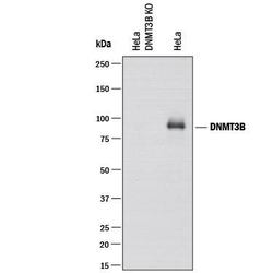
- Western Blot Shows Human DNMT3B Specificity by Using Knockout Cell Line. Western blot shows lysates of DNMT3B knockout HeLa cell line (KO) and HeLa human cervical epithelial carcinoma parental cell line. PVDF membrane was probed with 0.5 µg/mL of Sheep Anti-Human DNMT3B Antigen Affinity-purified Polyclonal Antibody (Catalog # AF7646) followed by HRP-conjugated Anti-Sheep IgG Secondary Antibody (Catalog # HAF016). A specific band was detected for DNMT3B at approximately 100-110 kDa (as indicated) in the parental HeLa cell line, but is not detectable in knockout HeLa cell line. GAPDH (Catalog # AF5718) is shown as a loading control. This experiment was conducted under reducing conditions and using Immunoblot Buffer Group 1.

- Submitted by
- R&D Systems (provider)
- Main image
- Experimental details
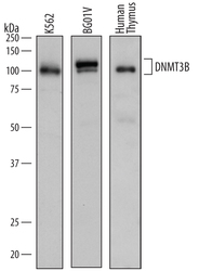
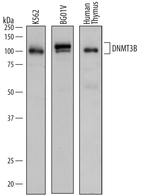
- Detection of Human DNMT3B by Western Blot. Western blot shows lysates of K562 human chronic myelogenous leukemia cell line, BG01V human embryonic stem cells, and human thymus tissue. PVDF membrane was probed with 0.2 µg/mL of Sheep Anti-Human DNMT3B Antigen Affinity-purified Polyclonal Antibody (Catalog # AF7646) followed by HRP-conjugated Anti-Sheep IgG Secondary Antibody (Catalog # HAF016). Specific bands were detected for DNMT3B at approximately 100-110 kDa (as indicated). This experiment was conducted under reducing conditions and using Immunoblot Buffer Group 1.
Supportive validation
- Submitted by
- R&D Systems (provider)
- Main image

- Experimental details
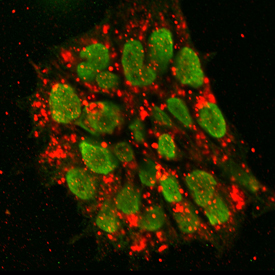
- DNMT3B in BG01V Human Embryonic Stem Cells. DNMT3B and SSEA-4 were detected in immersion fixed BG01V human embryonic stem cells. DNMT3B was detected using Sheep Anti-Human DNMT3B Antigen Affinity-purified Polyclonal Antibody (Catalog # AF7646) at 10 µg/mL for 3 hours at room temperature. Cells were stained using the NorthernLights™ 493-conjugated Anti-Sheep IgG Secondary Antibody (green; Catalog # NL012). SSEA-4 was detected using Mouse Anti-Human/Mouse SSEA-4 Monoclonal Antibody (Catalog # MAB1435) and stained using the NorthernLights™ 557-conjugated Anti-Mouse IgG Secondary Antibody (red; Catalog # NL007). Specific staining of DNMT3B was localized to nuclei. View our protocol for Fluorescent ICC Staining of Cells on Coverslips.