Explore
 Validate
 Learn
Learn Western blot
Antibody data
- Antibody Data
- Antigen structure
- References [3]
- Comments [0]
- Validations
- Western blot [5]
- Immunocytochemistry [1]
- Immunoprecipitation [1]
- Immunohistochemistry [1]
Submit
Validation data
Reference
Comment
Report error
- Product number
- GTX101316 - Provider product page

- Provider
- GeneTex
- Proper citation
- GeneTex Cat#GTX101316, RRID:AB_1240642
- Product name
- C3 antibody [C3], C-term
- Antibody type
- Polyclonal
- Reactivity
- Human, Mouse
- Host
- Rabbit
Submitted references Improved capillary electrophoresis method for the analysis of carbohydrate-deficient transferrin in human serum, avoiding interference by complement C3.
Human skin mast cells express complement factors C3 and C5.
Obesity-associated autoantibody production requires AIM to retain the immunoglobulin M immune complex on follicular dendritic cells.
Kuroda Y, Hamaguchi R, Moriyama K, Tanimoto T, Haginaka J
Journal of pharmaceutical and biomedical analysis 2013 Mar 25;76:81-6
Journal of pharmaceutical and biomedical analysis 2013 Mar 25;76:81-6
Human skin mast cells express complement factors C3 and C5.
Fukuoka Y, Hite MR, Dellinger AL, Schwartz LB
Journal of immunology (Baltimore, Md. : 1950) 2013 Aug 15;191(4):1827-34
Journal of immunology (Baltimore, Md. : 1950) 2013 Aug 15;191(4):1827-34
Obesity-associated autoantibody production requires AIM to retain the immunoglobulin M immune complex on follicular dendritic cells.
Arai S, Maehara N, Iwamura Y, Honda S, Nakashima K, Kai T, Ogishi M, Morita K, Kurokawa J, Mori M, Motoi Y, Miyake K, Matsuhashi N, Yamamura K, Ohara O, Shibuya A, Wakeland EK, Li QZ, Miyazaki T
Cell reports 2013 Apr 25;3(4):1187-98
Cell reports 2013 Apr 25;3(4):1187-98
No comments: Submit comment
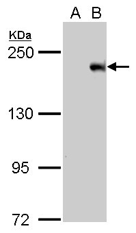
Supportive validation
- Submitted by
- GeneTex (provider)
- Main image

- Experimental details
- C3 antibody detects C3 protein by western blot analysis.A. 30 ?g 293T whole cell lysate/extractB. 30 ?g whole cell lysate/extract of human C3-transfected 293T cells5 % SDS-PAGEC3 antibody (GTX101316) dilution: 1:20000
- Validation comment
- WB
- Submitted by
- GeneTex (provider)
- Main image
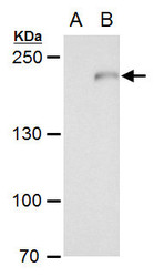
- Experimental details
- C3 antibody [C3], C-term detects C3 protein by western blot analysis.A. 30 ?g 293T whole cell extract B. 30 ?g whole cell extract of human C3-transfected 293T cells5 % SDS-PAGEC3 antibody [C3], C-term (GTX101316) dilution: 1:20000
- Validation comment
- WB

- Submitted by
- GeneTex (provider)
- Main image
- Experimental details
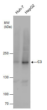
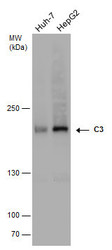
- C3 antibody detects C3 protein by western blot analysis. Various whole cell extracts (30 ?g) were separated by 5% SDS-PAGE, and the membrane was blotted with C3 antibody (GTX101316) diluted by 1:1000.
- Validation comment
- WB
- Submitted by
- GeneTex (provider)
- Main image
- Experimental details
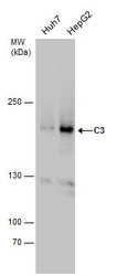
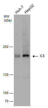
- C3 antibody detects C3 protein by western blot analysis. Various whole cell extracts (30 ?g) were separated by 5% SDS-PAGE, and the membrane was blotted with C3 antibody (GTX101316) diluted at a dilution of 1:1000.
- Validation comment
- WB
- Submitted by
- GeneTex (provider)
- Main image
- Experimental details
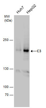
- C3 antibody detects C3 protein by western blot analysis. Various whole cell extracts (30 ?g) were separated by 5% SDS-PAGE, and the membrane was blotted with C3 antibody (GTX101316) diluted at a dilution of 1:1000. The HRP-conjugated anti-rabbit IgG antibody (GTX213110-01) was used to detect the primary antibody.
Supportive validation
- Submitted by
- GeneTex (provider)
- Main image

- Experimental details
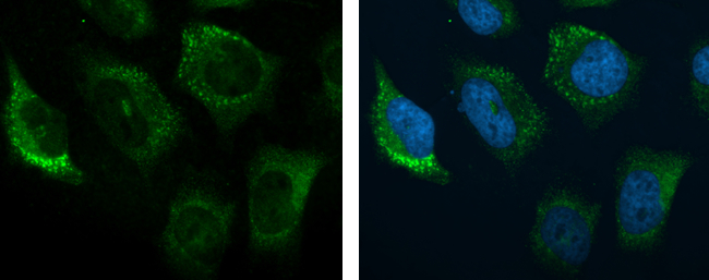
- C3 antibody [C3], C-term detects C3 protein at cytoplasm by immunofluorescent analysis.Sample: HeLa cells were fixed in 4% paraformaldehyde at RT for 15 min.Green: C3 protein stained by C3 antibody [C3], C-term (GTX101316) diluted at 1:200.Blue: Hoechst 33342 staining.
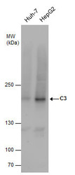
Supportive validation
- Submitted by
- GeneTex (provider)
- Main image

- Experimental details
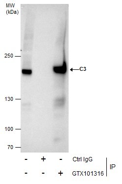
- Immunoprecipitation of C3 protein from HepG2 whole cell extracts using 5 £gg of C3 antibody [C3], C-term (GTX101316).Western blot analysis was performed using C3 antibody [C3], C-term (GTX101316).EasyBlot anti-Rabbit IgG (GTX221666-01) was used as a secondary reagent.
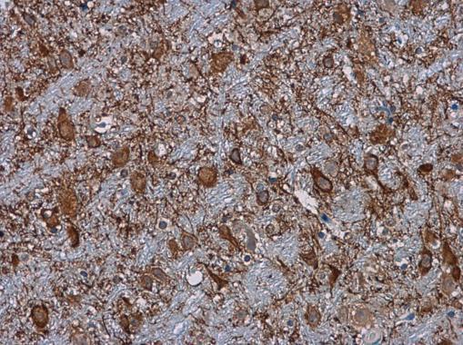
Supportive validation
- Submitted by
- GeneTex (provider)
- Main image

- Experimental details
- C3 antibody [C3], C-term detects C3 protein at cytoplasm in mouse brain by immunohistochemical analysis. Sample: Paraffin-embedded mouse brain. C3 antibody [C3], C-term (GTX101316) diluted at 1:500.