Explore
 Validate
 Learn
Learn Western blot
 ELISA
ELISA Immunoprecipitation
Antibody data
- Antibody Data
- Antigen structure
- References [0]
- Comments [0]
- Validations
- Western blot [4]
- Immunocytochemistry [1]
- Other assay [1]
Submit
Validation data
Reference
Comment
Report error
- Product number
- PA5-80959 - Provider product page

- Provider
- Invitrogen Antibodies
- Product name
- PTPN2 Polyclonal Antibody
- Antibody type
- Polyclonal
- Antigen
- Recombinant full-length protein
- Reactivity
- Human
- Host
- Rabbit
- Isotype
- IgG
- Vial size
- 100 µL
- Concentration
- 1 mg/mL
- Storage
- Store at 4°C short term. For long term storage, store at -20°C, avoiding freeze/thaw cycles.
No comments: Submit comment
Supportive validation
- Submitted by
- Invitrogen Antibodies (provider)
- Main image

- Experimental details
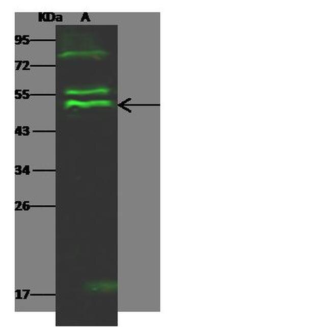
- Western blot analysis of PTPN2 in Lane A: MOLT4 Whole Cell Lysate (30 µg). Samples were probed using a PTPN2 Polyclonal Antibody (Product # PA5-80959) at a 1:500 dilution, followed by a Goat Anti-Rabbit IgG (H+L), Dylight 800 Secondary Antibody at a 1:10000 dilution. Western blot was performed under reducing conditions. Predicted band size:48 kDa. Observed band size:51 kDa.
- Submitted by
- Invitrogen Antibodies (provider)
- Main image
- Experimental details
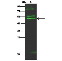
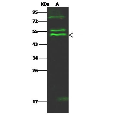
- Western Blot using PTPN2 Polyclonal Antibody (Product # PA5-80959) at 1:500 dilution. Lane A: MOLT4 Whole Cell Lysate. Lysates/proteins at 30 μg per lane. Secondary Goat Anti- RabbitIgG H&L (DyLight™ 800) at 1:10,000 dilution. Developed using the Odyssey technique. Performed under reducing conditions. Predicted band size: 48 kDa. Observed band size: 51 kDa. (We are unsure of the identity of these extra bands).
- Submitted by
- Invitrogen Antibodies (provider)
- Main image

- Experimental details
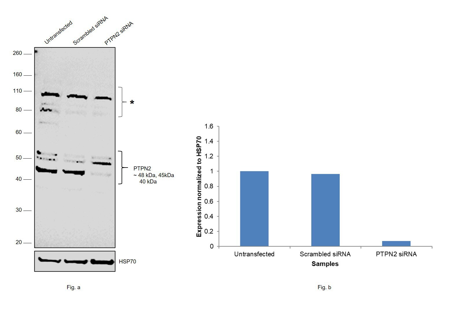
- Knockdown of PTPN2 was achieved by transfecting K-562 with PTPN2 specific siRNAs (Silencer® select Product # s11510, s11509). Western blot analysis (Fig. a) was performed using whole cell extracts from the PTPN2 knockdown cells (lane 3), non-targeting scrambled siRNA transfected cells (lane 2) and untransfected cells (lane 1). The blot was probed with PTPN2 Polyclonal Antibody (Product # PA5-80959, 1:1000 dilution) and Goat anti-Rabbit IgG (H+L) Superclonal™ Recombinant Secondary Antibody, HRP (Product # A27036, 1:10000 dilution). Densitometric analysis of this western blot for the 45 kDa isoform is shown in histogram (Fig. b). Decrease in signal upon siRNA mediated knock down confirms that antibody is specific to PTPN2.
- Submitted by
- Invitrogen Antibodies (provider)
- Main image
- Experimental details
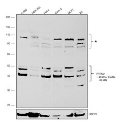
- Western blot was performed using Anti-PTPN2 Polyclonal Antibody (Product # PA5-80959) and 48 kDa, 45 kDa, and 40kDa bands corresponding to PTPN2 (Tyrosine-protein phosphatase non-receptor type 2) was observed across cell lines tested along with uncharacterized bands at ~80 kDa and 110 kDa. Whole cell extracts (30 µg lysate) of K-562 (Lane 1), HEK-293 (Lane 2), HeLa (Lane 3), Caco-2 (Lane 4), MCF7 (Lane 5) and BJ (Lane 6) were electrophoresed using NuPAGE™ 10% Bis-Tris Protein Gel (Product # NP0301BOX). Resolved proteins were then transferred onto a nitrocellulose membrane (Product # IB23001) by iBlot® 2 Dry Blotting System (Product # IB21001). The blot was probed with the primary antibody (1:1000 dilution) and detected by chemiluminescence with Goat anti-Rabbit IgG (H+L) Superclonal™ Recombinant Secondary Antibody, HRP (Product # A27036, 1:10000 dilution) using the iBright FL 1000 (Product # A32752). Chemiluminescent detection was performed using Novex® ECL Chemiluminescent Substrate Reagent Kit (Product # WP20005).
Supportive validation
- Submitted by
- Invitrogen Antibodies (provider)
- Main image
- Experimental details
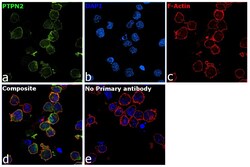
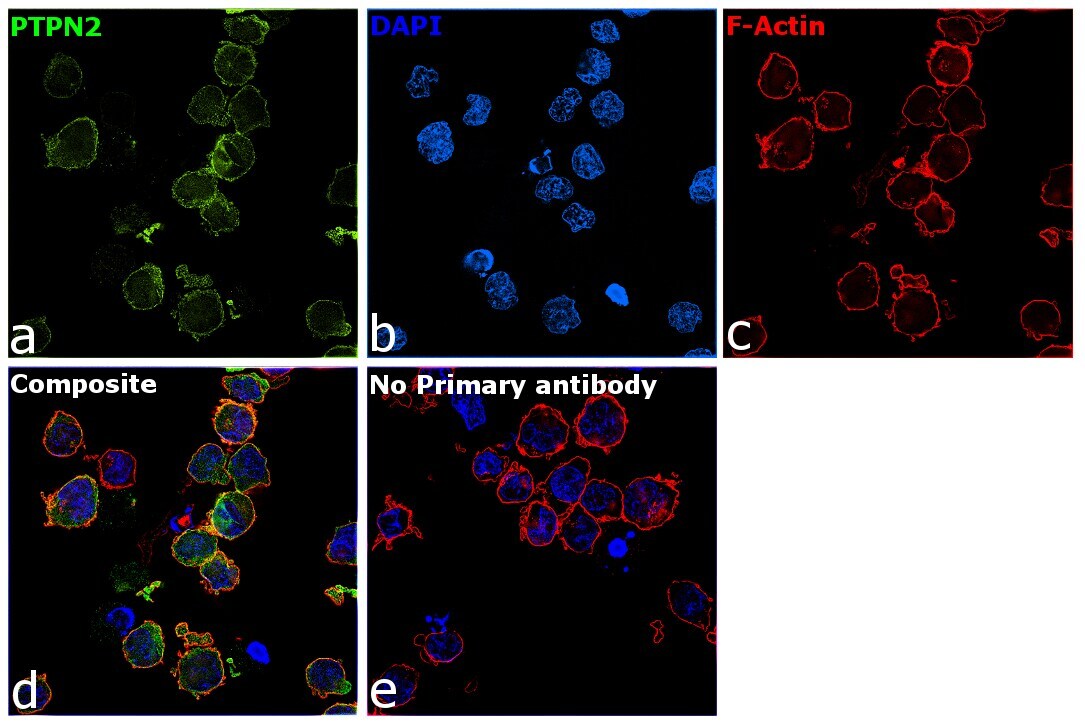
- Immunofluorescence analysis of Tyrosine-protein phosphatase non-receptor type 2 was performed using 70% confluent log phase K-562 cells. The cells were fixed with 4% paraformaldehyde for 10 minutes, permeabilized with 0.1% Triton™ X-100 for 10 minutes, and blocked with 2% BSA for 45 minutes at room temperature. The cells were labeled with PTPN2 Polyclonal Antibody (Product # PA5-80959) at 1:100 in 0.1% BSA, incubated at 4 degree celsius overnight and then labeled with Donkey anti-Rabbit IgG (H+L) Highly Cross-Adsorbed Secondary Antibody, Alexa Fluor Plus 488 (Product # A32790), (1:2500 dilution), for 45 minutes at room temperature (Panel a: Green). Nuclei (Panel b:Blue) were stained with ProLong™ Diamond Antifade Mountant with DAPI (Product # P36962). F-actin (Panel c: Red) was stained with Rhodamine Phalloidin (Product # R415, 1:300). Panel d represents the merged image showing cytoplasmic and membrane localization. Panel e represents control cells with no primary antibody to assess background. The images were captured at __21 magnification.
Supportive validation
- Submitted by
- Invitrogen Antibodies (provider)
- Main image

- Experimental details
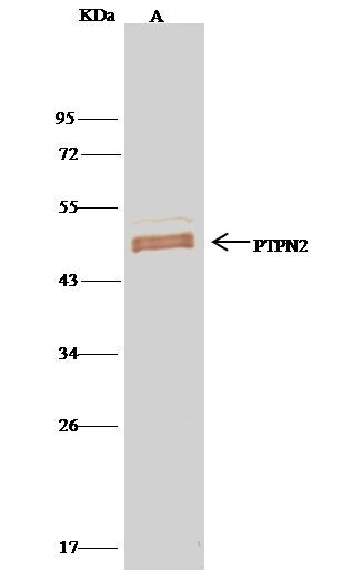
- PTPN2 Immunoprecipitation using: Lane A: 0.5 mg MOLT-4 Whole Cell Lysate 1 µL with PTPN2 Polyclonal Antibody (Product # PA5-80959) and 15 µL of 50 % Protein G agarose. Primary antibody: PTPN2 Polyclonal Antibody, at 1:500 dilution. Secondary antibody: Clean-Bloto IP Detection Reagent (HRP) at 1:500 dilution. Developed using the DAB staining technique. Performed under reducing conditions. Predicted band size: 48 kDa. Observed band size: 48 kDa.