Explore
 Validate
 Learn
Learn Western blot
Antibody data
- Antibody Data
- Antigen structure
- References [3]
- Comments [0]
- Validations
- Western blot [6]
- Immunocytochemistry [2]
- Immunohistochemistry [1]
- Other assay [2]
Submit
Validation data
Reference
Comment
Report error
- Product number
- PA5-27384 - Provider product page

- Provider
- Invitrogen Antibodies
- Product name
- Blooms Syndrome Polyclonal Antibody
- Antibody type
- Polyclonal
- Antigen
- Recombinant protein fragment
- Reactivity
- Human
- Host
- Rabbit
- Isotype
- IgG
- Vial size
- 100 µL
- Concentration
- 1.34 mg/mL
- Storage
- Store at 4°C short term. For long term storage, store at -20°C, avoiding freeze/thaw cycles.
Submitted references Resolution of ROS-induced G-quadruplexes and R-loops at transcriptionally active sites is dependent on BLM helicase.
Functional conservation of RecQ helicase BLM between humans and Drosophila melanogaster.
Analysis of alternative lengthening of telomere markers in BRCA1 defective cells.
Tan J, Wang X, Phoon L, Yang H, Lan L
FEBS letters 2020 May;594(9):1359-1367
FEBS letters 2020 May;594(9):1359-1367
Functional conservation of RecQ helicase BLM between humans and Drosophila melanogaster.
Cox RL, Hofley CM, Tatapudy P, Patel RK, Dayani Y, Betcher M, LaRocque JR
Scientific reports 2019 Nov 26;9(1):17527
Scientific reports 2019 Nov 26;9(1):17527
Analysis of alternative lengthening of telomere markers in BRCA1 defective cells.
Kargaran PK, Yasaei H, Anjomani-Virmouni S, Mangiapane G, Slijepcevic P
Genes, chromosomes & cancer 2016 Nov;55(11):864-76
Genes, chromosomes & cancer 2016 Nov;55(11):864-76
No comments: Submit comment
Supportive validation
- Submitted by
- Invitrogen Antibodies (provider)
- Main image

- Experimental details
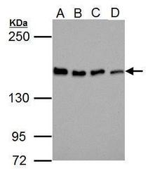
- Western blot analysis of BLM using 30 µg of A) Jurkat (B) Raji (C) K562 and D) NCI-H929 lysate. Samples were loaded onto a 5% SDS-PAGE gel and probed with a BLM polyclonal antibody (Product # PA5-27384) at a dilution of 1:1000.
- Submitted by
- Invitrogen Antibodies (provider)
- Main image
- Experimental details
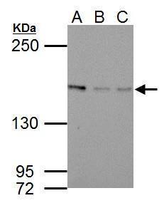
- Western Blot using Blooms Syndrome Polyclonal Antibody (Product # PA5-27384). Sample (30 µg of whole cell lysate). A: Jurkat. B: Raji. C: K562. D: NCI-H929. 5% SDS PAGE. Blooms Syndrome Polyclonal Antibody (Product # PA5-27384) diluted at 1:1,000. The HRP-conjugated anti-rabbit IgG antibody was used to detect the primary antibody.
- Submitted by
- Invitrogen Antibodies (provider)
- Main image
- Experimental details
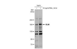
- Western Blot using Blooms Syndrome Polyclonal Antibody (Product # PA5-27384). Untreated (–) and treated (+) THP-1 cells were untreated or treated with 10 ng/mL PMA for 24 hrs (30 µg) were separated by 5% SDS-PAGE, and the membrane was blotted with Blooms Syndrome Polyclonal Antibody (Product # PA5-27384) diluted at 1:1,000. The HRP-conjugated anti-rabbit IgG antibody was used to detect the primary antibody, and the signal was developed with Trident ECL plus-Enhanced.
- Submitted by
- Invitrogen Antibodies (provider)
- Main image
- Experimental details
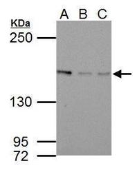
- Western Blot using Blooms Syndrome Polyclonal Antibody (Product # PA5-27384). Sample (30 µg of whole cell lysate). A: NIH-3T3. B: JC. C: BCL-1. 5% SDS PAGE. Blooms Syndrome Polyclonal Antibody (Product # PA5-27384) diluted at 1:1,000. The HRP-conjugated anti-rabbit IgG antibody was used to detect the primary antibody.
- Submitted by
- Invitrogen Antibodies (provider)
- Main image
- Experimental details
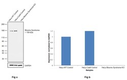
- Knockout of Blooms syndrome was achieved by CRISPR-Cas9 genome editing using LentiArray™ Lentiviral sgRNA (Product # A32042, Assay ID CRISPR815029_LV) and LentiArray Cas9 Lentivirus (Product # A32064). Western blot analysis of Blooms syndrome was performed by loading 30 µg of HeLa Wild type (Lane 1), HeLa Cas9 (Lane 2) and HeLa Blooms syndrome KO (Lane 3) modified whole cell extracts. The samples were electrophoresed using NuPAGE™ Novex™ 4-12% Bis-Tris Protein Gel (Product # NP0322BOX). Resolved proteins were then transferred onto a nitrocellulose membrane (Product # IB23001) by iBlot® 2 Dry Blotting System (Product # IB21001). The blot was probed with Anti-Blooms Syndrome Polyclonal Antibody (Product # PA5-27384, 1:1,000 dilution) and Goat anti-Rabbit IgG (H+L) Superclonal™ Recombinant Secondary Antibody, HRP (Product # A27036, 1:5,000 dilution) using the iBright FL 1000 (Product # A32752). Chemiluminescent detection was performed using SuperSignal™ West Dura Extended Duration Substrate (Product # 34076). Loss of signal upon CRISPR mediated knockout (KO) using the LentiArray™ CRISPR product line confirms that antibody is specific to Blooms syndrome.
- Submitted by
- Invitrogen Antibodies (provider)
- Main image
- Experimental details
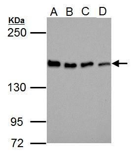
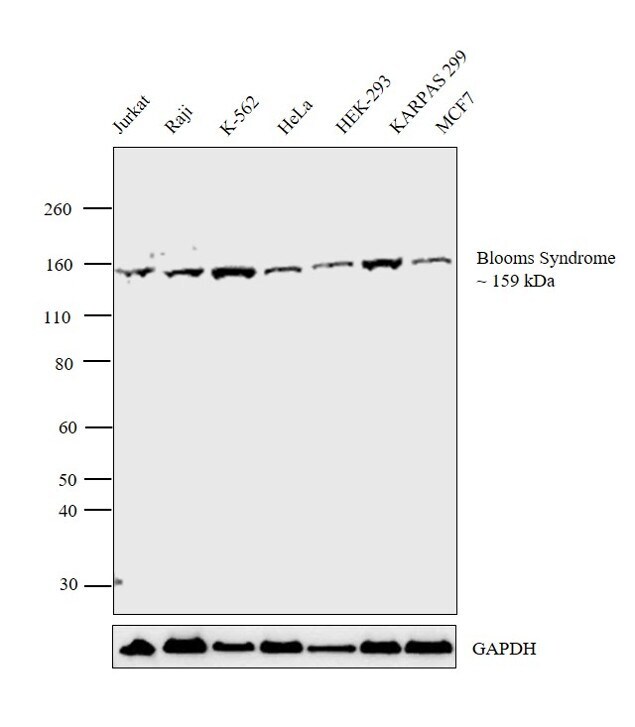
- Western blot analysis was performed on modified whole cell extracts (1% SDS) (30 µg lysate) of Jurkat (Lane 1), Raji (Lane 2), K-562 (Lane 3), HeLa (Lane 4), HEK-293 (Lane 5), KARPAS 299 (Lane 6) and MCF7 (Lane 7). The blot was probed with Anti- Blooms Syndrome Polyclonal Antibody (Product # PA5-27384, 1:1000 dilution) and detected by chemiluminescence using Goat anti Rabbit IgG (H+L) Superclonal™ Secondary Antibody, HRP conjugate (Product # A27036, 0.25 µg/mL, 1:4000 dilution). A 159 kDa band corresponding to Blooms Syndrome was detected across the cell lines tested.
Supportive validation
- Submitted by
- Invitrogen Antibodies (provider)
- Main image
- Experimental details
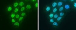
- Immunocytochemistry-Immunofluorescence analysis of Blooms Syndrome was performed in A431 cells fixed in 4% paraformaldehyde at RT for 15 min. Green: Blooms Syndrome Polyclonal Antibody (Product # PA5 27384) diluted at 1:200. Blue: Hoechst 33342 staining.
- Submitted by
- Invitrogen Antibodies (provider)
- Main image
- Experimental details
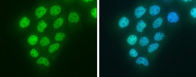
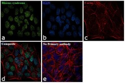
- Immunofluorescence analysis of Blooms Syndrome was performed using 70% confluent log phase A-431 cells. The cells were fixed with 4% paraformaldehyde for 10 minutes, permeabilized with 0.1% Triton™ X-100 for 15 minutes, and blocked with 1% BSA for 1 hour at room temperature. The cells were labeled with Blooms Syndrome Polyclonal Antibody (Product # PA5-27384) at 5µg/mL in 0.1% BSA, incubated at 4 degree Celsius overnight and then labeled with Goat anti-Rabbit IgG (H+L) Superclonal™ Secondary Antibody, Alexa Fluor® 488 conjugate (Product # A27034) at a dilution of 1:2000 for 45 minutes at room temperature (Panel a: green). Nuclei (Panel b: blue) were stained with ProLong™ Diamond Antifade Mountant with DAPI (Product # P36962). F-actin (Panel c: red) was stained with Rhodamine Phalloidin (Product # R415). Panel d represents the merged image showing Nuclear localization. Panel e represents control cells with no primary antibody to assess background. The images were captured at 60X magnification.

Supportive validation
- Submitted by
- Invitrogen Antibodies (provider)
- Main image
- Experimental details
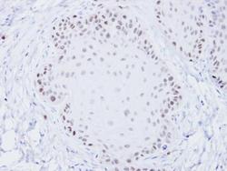
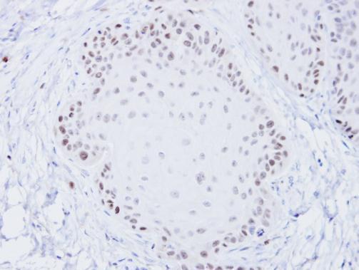
- Immunohistochemical analysis of paraffin-embedded Cal27 xenograft, using BLM (Product # PA5-27384) antibody at 1:100 dilution. Antigen Retrieval: EDTA based buffer, pH 8.0, 15 min.
Supportive validation
- Submitted by
- Invitrogen Antibodies (provider)
- Main image

- Experimental details
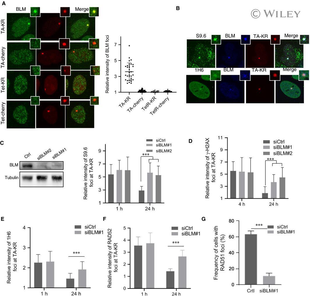
- BLM is required for R-loop and G-quadruplex resolution. (A) U2OS TRE cells transfected with TA-KR/TA-Cherry/tetR-KR/tetR-Cherry plasmids were exposed to light for 30 min for KR activation and allowed to recover for 30 min before harvest. Cells were stained with BLM Ab. For quantification of A to F, mean values of intensity with SD from 30 cells in three independent experiments are given. P -value is calculated by unpaired t -test, *** P < 0.001. (B) The colocalization of S9.6 or 1H6 with BLM at TA-KR sites in cells with the same treatment as 3A. (C-D) U2OS TRE cells transfected with siBLM#1 or siBLM#2, and TA-KR were exposed to light for 30 min for KR activation and allowed to recover for indicated time. Relative intensity of S9.6 (C) and gammaH2AX (D) via Abs at sites of TA-KR is quantified. WB of BLM and Tubulin in indicated cell lysates are shown. (E) U2OS TRE cells transfected with siBLM#1 and TA-KR were exposed to light for 30 min for KR activation and allowed to recover for indicated time. Relative intensity of 1H6 via Abs at sites of TA-KR is quantified. (F) U2OS TRE cells transfected with siBLM, TA-KR, and GFP-RAD52 were exposed to light for 30 min for KR activation and allowed to recover for indicated time. Relative intensity of GFP-RAD52 at sites of TA-KR is quantified. (G) U2OS TRE cells transfected with siBLM#1 and TA-KR were exposed to light for 30 min for KR activation and allowed to recover for 24 h. The frequency of RAD51 via Ab at sites of TA-KR is quantified
- Submitted by
- Invitrogen Antibodies (provider)
- Main image

- Experimental details
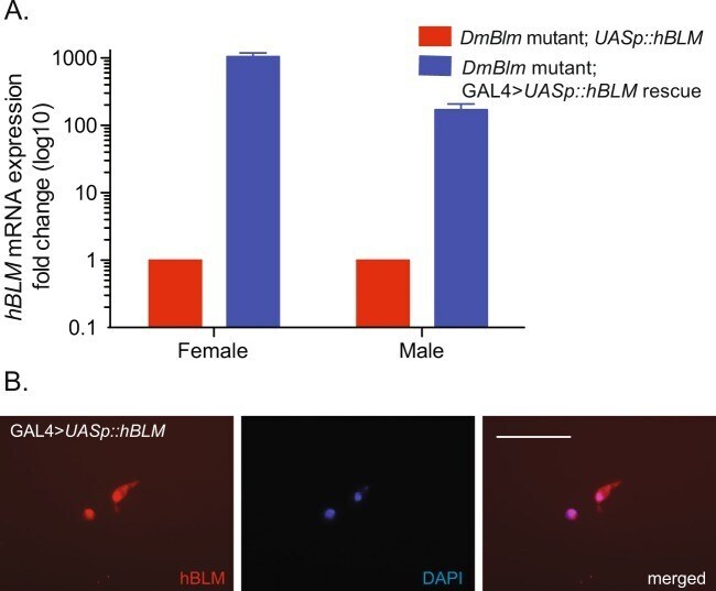
- Figure 3 hBLM expression and localization. ( A ) Flies with Act5c::GAL4 and UASp::hBLM transgenes (blue) showed greater hBLM mRNA expression than baseline levels without the GAL4 > UASp expression system (red). Mean fold change and standard errors of the mean are shown. ( B ) UASp::hBLM was co-transfected with Act5c::GAL4 into S2 Drosophila cells. Localizing to the nucleus (stained with DAPI) was evident in transfected cells using hBLM-specific immunofluorescence. Scale bar is 32 um.