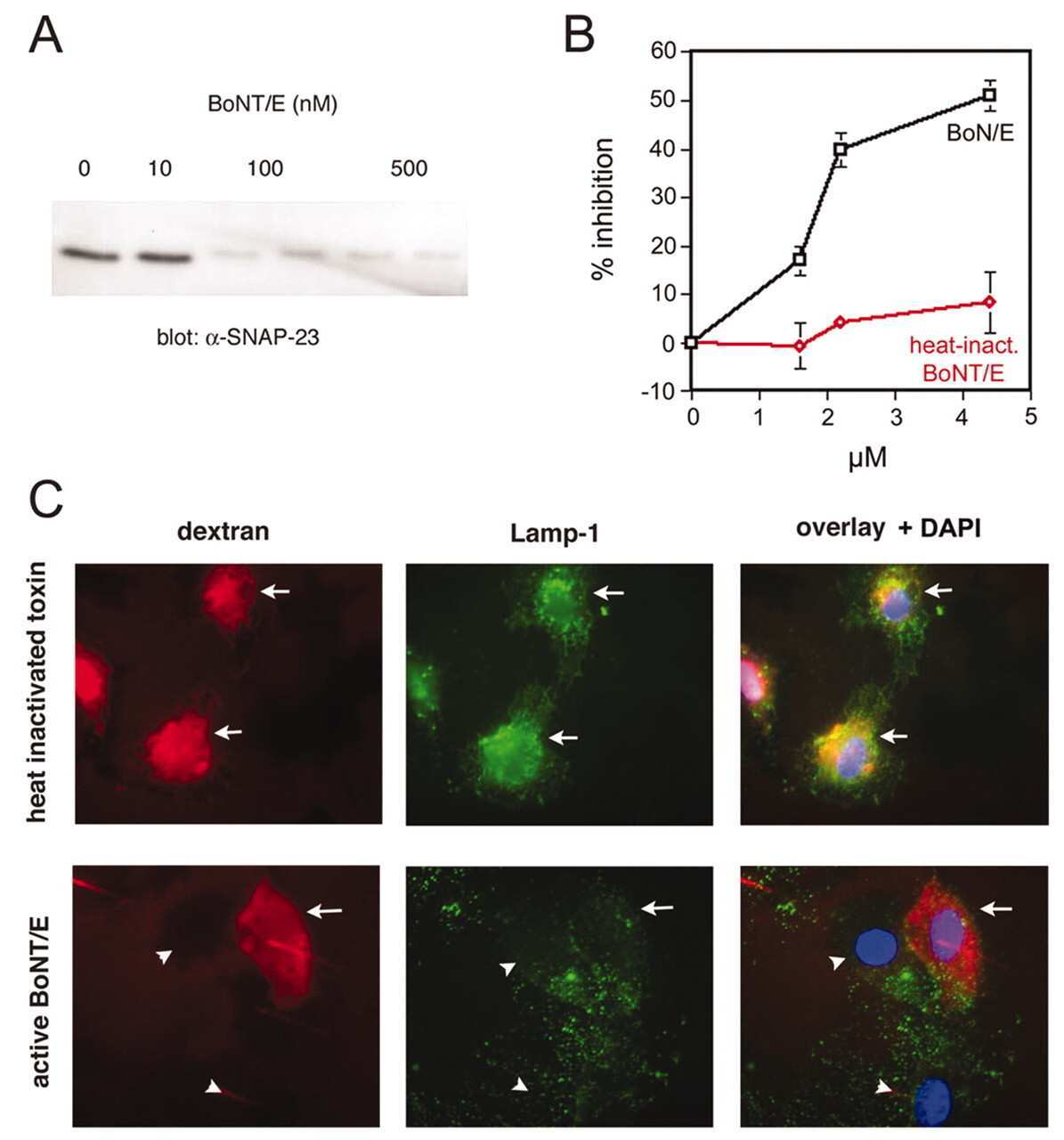
Explore
 Validate
 Learn
Learn Western blot
Antibody data
- Antibody Data
- Antigen structure
- References [4]
- Comments [0]
- Validations
- Western blot [4]
- Immunocytochemistry [1]
- Other assay [3]
Submit
Validation data
Reference
Comment
Report error
- Product number
- PA1-738 - Provider product page

- Provider
- Invitrogen Antibodies
- Product name
- SNAP23 Polyclonal Antibody
- Antibody type
- Polyclonal
- Antigen
- Synthetic peptide
- Description
- PA1-738 detects synaptosomal-associated protein 23 (SNAP-23) from human, mouse and rat samples.
- Concentration
- 1 mg/mL
Submitted references Stimulus-induced S-nitrosylation of Syntaxin 4 impacts insulin granule exocytosis.
Developmental changes in the milk fat globule membrane proteome during the transition from colostrum to milk.
Effects of endurance exercise training on insulin signaling in human skeletal muscle: interactions at the level of phosphatidylinositol 3-kinase, Akt, and AS160.
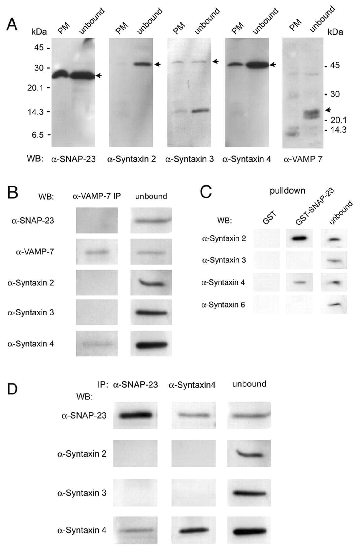
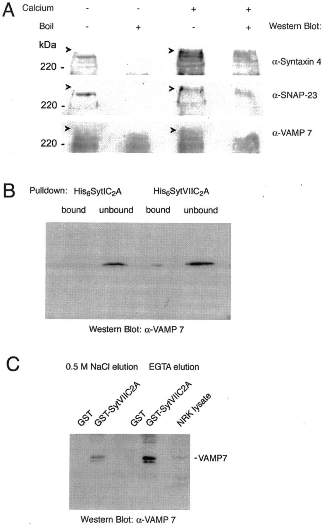
Identification of SNAREs involved in synaptotagmin VII-regulated lysosomal exocytosis.
Wiseman DA, Kalwat MA, Thurmond DC
The Journal of biological chemistry 2011 May 6;286(18):16344-54
The Journal of biological chemistry 2011 May 6;286(18):16344-54
Developmental changes in the milk fat globule membrane proteome during the transition from colostrum to milk.
Reinhardt TA, Lippolis JD
Journal of dairy science 2008 Jun;91(6):2307-18
Journal of dairy science 2008 Jun;91(6):2307-18
Effects of endurance exercise training on insulin signaling in human skeletal muscle: interactions at the level of phosphatidylinositol 3-kinase, Akt, and AS160.
Frøsig C, Rose AJ, Treebak JT, Kiens B, Richter EA, Wojtaszewski JF
Diabetes 2007 Aug;56(8):2093-102
Diabetes 2007 Aug;56(8):2093-102
Identification of SNAREs involved in synaptotagmin VII-regulated lysosomal exocytosis.
Rao SK, Huynh C, Proux-Gillardeaux V, Galli T, Andrews NW
The Journal of biological chemistry 2004 May 7;279(19):20471-9
The Journal of biological chemistry 2004 May 7;279(19):20471-9
No comments: Submit comment
Supportive validation
- Submitted by
- Invitrogen Antibodies (provider)
- Main image

- Experimental details
- Western blot of SNAP-23 on rat brain protein extract using Product # PA1-738.
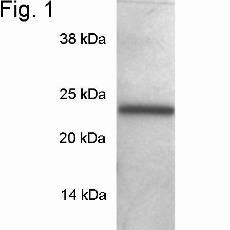
- Submitted by
- Invitrogen Antibodies (provider)
- Main image

- Experimental details
- Western blot of SNAP-23 on rat brain protein extract using Product # PA1-738.

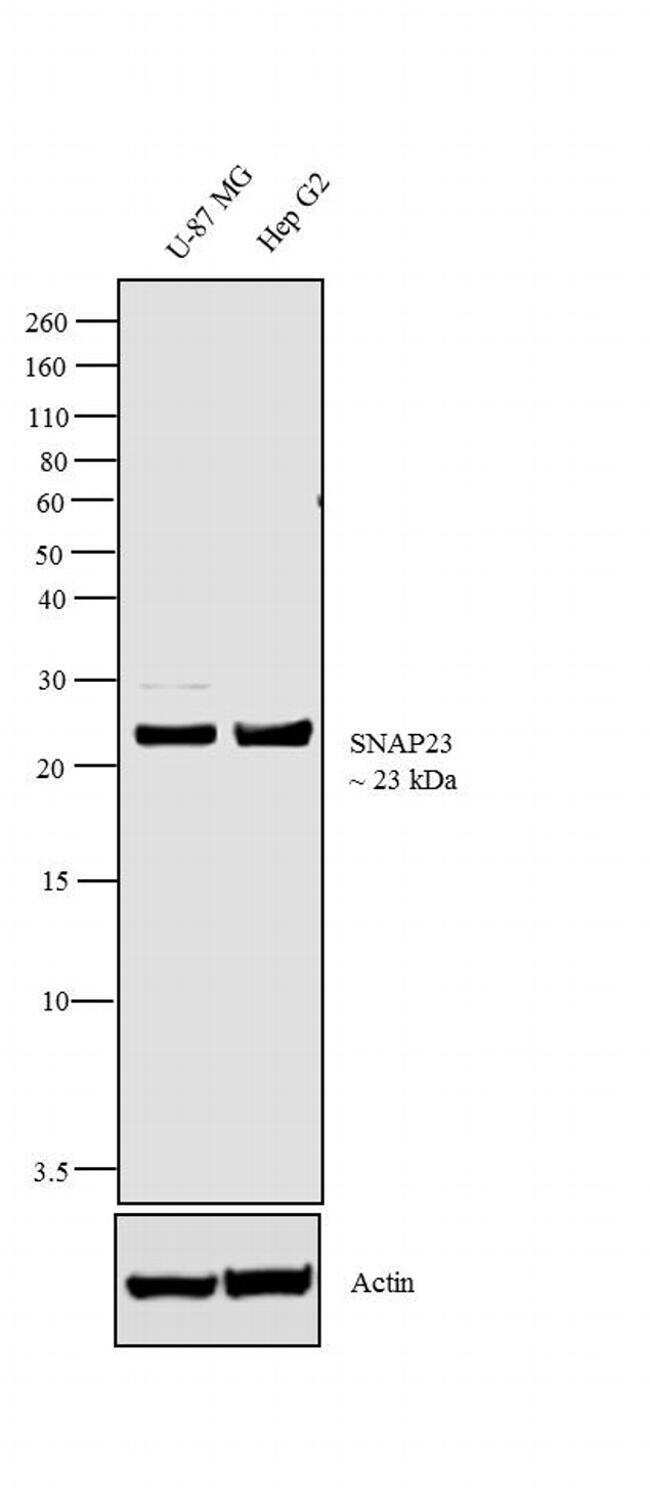
- Submitted by
- Invitrogen Antibodies (provider)
- Main image

- Experimental details
- Western blot analysis was performed on membrane enriched extracts (30 µg lysate) of U-87 MG (Lane 1) and Hep G2 (Lane 2). The blot was probed with Anti-SNAP23 Rabbit Polyclonal Antibody (Product # PA1-738, 2 µg/mL) and detected by chemiluminescence using Goat anti-Rabbit IgG (H+L) Superclonal™ Secondary Antibody, HRP conjugate (Product # A27036, 0.4 µg/mL, 1:2500 dilution). A 23 kDa band corresponding to SNAP23 was observed across the cell lines tested. Known quantity of protein samples were electrophoresed using Novex® NuPAGE® 12 % Bis-Tris gel (Product # NP0342BOX), XCell SureLock™ Electrophoresis System (Product # EI0002) and Novex® Sharp Pre-Stained Protein Standard (Product # LC5800). Resolved proteins were then transferred onto a nitrocellulose membrane with iBlot® 2 Dry Blotting System (Product # IB21001). The membrane was probed with the relevant primary and secondary Antibody following blocking with 5 % skimmed milk. Chemiluminescent detection was performed using Pierce™ ECL Western Blotting Substrate (Product # 32106).
- Submitted by
- Invitrogen Antibodies (provider)
- Main image
- Experimental details
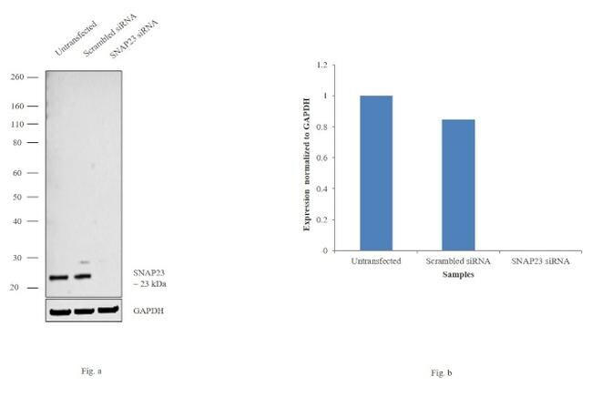
- Knockdown of SNAP23 was achieved by transfecting U87MG cells with SNAP23 specific siRNAs (Silencer® select Product # s16709,s16710). Western blot analysis (Fig. a) was performed using membrane enriched extracts from the SNAP23 knockdown cells (lane 3), non-specific scrambled siRNA transfected cells (lane 2) and untransfected cells (lane 1). The blot was probed with SNAP23 Polyclonal Antibody (Product # PA1-738, 1µg/ml) and Goat anti-Rabbit IgG (H+L) Superclonal™ Secondary Antibody, HRP conjugate (Product # A27036, 0.25µg/ml, 1:4000 dilution). Densitometric analysis of this western blot is shown in histogram (Fig. b). Decrease in signal upon siRNA mediated knock down confirms that antibody is specific to SNAP23.
Supportive validation
- Submitted by
- Invitrogen Antibodies (provider)
- Main image
- Experimental details
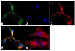
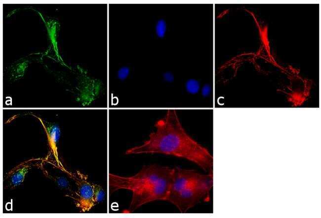
- Immunofluorescence analysis of SNAP-23 was performed using 90% confluent log phase U-87 MG cells. The cells were fixed with 4% paraformaldehyde for 10 minutes, permeabilized with 0.1% Triton™ X-100 for 10 minutes, and blocked with 1% BSA for 1 hour at room temperature. The cells were labeled with SNAP23 Rabbit Polyclonal Antibody (Product # PA1-738) at 2µg/mL in 0.1% BSA and incubated for 3 hours at room temperature and then labeled with Goat anti-Rabbit IgG (H+L) Superclonal™ Secondary Antibody, Alexa Fluor® 488 conjugate (Product # A27034) at a dilution of 1:2000 for 45 minutes at room temperature (Panel a: green). Nuclei (Panel b: blue) were stained with SlowFade® Gold Antifade Mountant with DAPI (Product # S36938). F-actin (Panel c: red) was stained with Alexa Fluor® 555 Rhodamine Phalloidin (Product # R415, 1:300). Panel d represents the merged image showing membrane localization. Panel e shows the no primary antibody control. The images were captured at 60X magnification.
Supportive validation
- Submitted by
- Invitrogen Antibodies (provider)
- Main image
- Experimental details
- NULL
- Submitted by
- Invitrogen Antibodies (provider)
- Main image

- Experimental details
- NULL
- Submitted by
- Invitrogen Antibodies (provider)
- Main image
- Experimental details
- NULL