 Explore
 Validate
 Learn
Learn Western blot
 Immunoprecipitation
Antibody data
- Antibody Data
- Antigen structure
- References [0]
- Comments [0]
- Validations
- Western blot [1]
- Immunocytochemistry [3]
- Flow cytometry [3]
Submit
Validation data
Reference
Comment
Report error
- Product number
- GTX80684 - Provider product page

- Provider
- GeneTex
- Proper citation
- GeneTex Cat#GTX80684, RRID:AB_625459
- Product name
- Dynein antibody [74.1]
- Antibody type
- Monoclonal
- Reactivity
- Human, Rat, Bovine, Porcine, Sheep
- Host
- Mouse
- Storage
- Keep as concentrated solution. Aliquot and store at -20?C or below. Avoid multiple freeze-thaw cycles.
No comments: Submit comment
Supportive validation
- Submitted by
- GeneTex (provider)
- Main image
- Experimental details
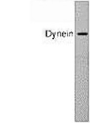
- Western blot of cytoplasmic dynein in HeLa cell lysate using GTX80684.

Supportive validation
- Submitted by
- GeneTex (provider)
- Main image
- Experimental details
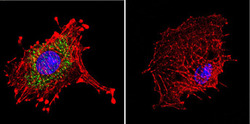
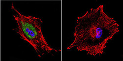
- Immunofluorescent analysis of Dynein in C6 Cells. Cells were grown on chamber slides and fixed with formaldehyde prior to staining. Cells were probed without (control) or with a Dynein monoclonal antibody (GTX80684) at a dilution of 1:20 overnight at 4 C, washed with PBS and incubated with a DyLight-488 conjugated secondary antibody. Dynein staining (green), F-Actin staining with Phalloidin (red) and nuclei with DAPI (blue) is shown.
- Submitted by
- GeneTex (provider)
- Main image

- Experimental details
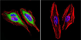
- Immunofluorescent analysis of Dynein in HeLa Cells. Cells were grown on chamber slides and fixed with formaldehyde prior to staining. Cells were probed without (control) or with a Dynein monoclonal antibody (GTX80684) at a dilution of 1:20 overnight at 4 C, washed with PBS and incubated with a DyLight-488 conjugated secondary antibody. Dynein staining (green), F-Actin staining with Phalloidin (red) and nuclei with DAPI (blue) is shown.
- Submitted by
- GeneTex (provider)
- Main image
- Experimental details
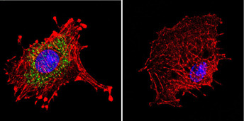
- Immunofluorescent analysis of Dynein in U87-MG Cells. Cells were grown on chamber slides and fixed with formaldehyde prior to staining. Cells were probed without (control) or with a Dynein monoclonal antibody (GTX80684) at a dilution of 1:20 overnight at 4 C, washed with PBS and incubated with a DyLight-488 conjugated secondary antibody. Dynein staining (green), F-Actin staining with Phalloidin (red) and nuclei with DAPI (blue) is shown.

Supportive validation
- Submitted by
- GeneTex (provider)
- Main image
- Experimental details
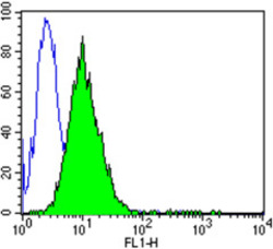
- Flow cytometry analysis of Dynein showing positive staining in the cytoplasm of C6 cells compared to an isotype control (blue). Cells were harvested, adjusted to a concentration of 1-5x10^6 cells/ml, fixed with 2% paraformaldehyde and washed with PBS. Cells were penetrated by dropping the supernatant, adding 90% methanol and incubated for 10 minutes at room temperature. Follwing penetration, cells were blocked with a 2% solution of BSA-PBS for 30 min at room temperature and incubated with a Dynein monoclonal antibody (GTX80684) at a dilution of 1 ug/test for 60 min at room temperature. Cells were then incubated for 40 min at room temperature in the dark using a Dylight 488-conjugated goat anti-mouse IgG (H+L) secondary antibody and re-suspended in PBS for FACS analysis.

- Submitted by
- GeneTex (provider)
- Main image
- Experimental details
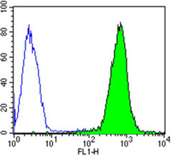
- Flow cytometry analysis of Dynein showing strong positive staining in the cytoplasm of Hela cells compared to an isotype control (blue). Cells were harvested, adjusted to a concentration of 1-5x10^6 cells/ml, fixed with 2% paraformaldehyde and washed with PBS. Cells were penetrated by dropping the supernatant, adding 90% methanol and incubated for 10 minutes at room temperature. Follwing penetration, cells were blocked with a 2% solution of BSA-PBS for 30 min at room temperature and incubated with a Dynein monoclonal antibody (GTX80684) at a dilution of 2 ug/test for 60 min at room temperature. Cells were then incubated for 40 min at room temperature in the dark using a Dylight 488-conjugated goat anti-mouse IgG (H+L) secondary antibody and re-suspended in PBS for FACS analysis.

- Submitted by
- GeneTex (provider)
- Main image
- Experimental details
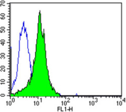
- Flow cytometry analysis of Dynein showing positive staining in the cytoplasm of U87-MG cells compared to an isotype control (blue). Cells were harvested, adjusted to a concentration of 1-5x10^6 cells/ml, fixed with 2% paraformaldehyde and washed with PBS. Cells were penetrated by dropping the supernatant, adding 90% methanol and incubated for 10 minutes at room temperature. Following penetration, cells were blocked with a 2% solution of BSA-PBS for 30 min at room temperature and incubated with a Dynein monoclonal antibody (GTX80684) at a dilution of 2 ug/test for 60 min at room temperature. Cells were then incubated for 40 min at room temperature in the dark using a Dylight 488-conjugated goat anti-mouse IgG (H+L) secondary antibody and re-suspended in PBS for FACS analysis.
