Antibody data
- Antibody Data
- Antigen structure
- References [1]
- Comments [0]
- Validations
- Western blot [3]
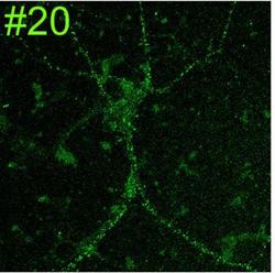
- Immunocytochemistry [2]
Submit
Validation data
Reference
Comment
Report error
- Product number
- 73-097 - Provider product page
- Provider
- Antibodies Incorporated / NeuroMab
- Product name
- Anti-GluN2B/NR2B glutamate receptor
- Antibody type
- Monoclonal
- Description
- TC Supernatant
- Reactivity
- Human, Mouse, Rat
- Host
- Mouse
- Conjugate
- Unconjugated
- Isotype
- IgG
- Antibody clone number
- N59/20
- Vial size
- 5 mL
- Concentration
- Lot dependent
- Storage
- Aliquot and store at -20 degrees Celsius
Submitted references Developmentally dynamic colocalization patterns of DSCAM with adhesion and synaptic proteins in the mouse retina.
de Andrade GB, Kunzelman L, Merrill MM, Fuerst PG
Molecular vision 2014;20:1422-33
No comments: Submit comment
Supportive validation
- Submitted by
- Antibodies Incorporated / NeuroMab (provider)
- Main image
- Experimental details
- Immunoblot against rat brain membranes (RBM) and neuronal lysates from NR2B knockout (KO) and heterozygote (Het) mice. Samples courtesy of Ben Hall and Anirvan Ghosh (UCSD).
- Submitted by
- Antibodies Incorporated / NeuroMab (provider)
- Main image
- Experimental details
- Immunoblots on brain membranes prepared from whole rat (RBM) and mouse (MBM) brain, and from human cerebral cortex [HBM(Cx)] and hippocampus [HBM(H)] probed with N59/20 (left) or N52A/42 (right) TC supe.
- Submitted by
- Antibodies Incorporated / NeuroMab (provider)
- Main image
- Experimental details
- Immunoblots on brain membranes prepared from whole rat.
Supportive validation
- Submitted by
- Antibodies Incorporated / NeuroMab (provider)
- Main image
- Experimental details
- Adult rat hippocampus immunofluorescence under standard protocol (left) and with antigen retrieval via pepsin pretreatment (right).
- Submitted by
- Antibodies Incorporated / NeuroMab (provider)
- Main image
- Experimental details
- Cultured rat hippocampal neuron immunofluorescence. Image courtesy of Anthone Dunah (Harvard) and Morgan Sheng (MIT).