Explore
 Validate
 Learn
Learn Western blot
Antibody data
- Antibody Data
- Antigen structure
- References [3]
- Comments [0]
- Validations
- Western blot [1]
Submit
Validation data
Reference
Comment
Report error
- Product number
- 600-401-905 - Provider product page

- Provider
- Rockland Immunochemicals, Inc.
- Proper citation
- Rockland Cat#600-401-905, RRID:AB_2096271
- Product name
- Anti-EGFR (RABBIT) Antibody - 600-401-905
- Antibody type
- Polyclonal
- Vial size
- 100 µl
Submitted references Oriented immobilization to nanoparticles enhanced the therapeutic efficacy of antibody drugs.
Therapeutic efficacy of a synthetic epsin mimetic peptide in glioma tumor model: uncovering multiple mechanisms beyond the VEGF-associated tumor angiogenesis.
EGFR-targeted Chimeras of Pseudomonas ToxA released into the extracellular milieu by attenuated Salmonella selectively kill tumor cells.
Iijima M, Araki K, Liu Q, Somiya M, Kuroda S
Acta biomaterialia 2019 Mar 1;86:373-380
Acta biomaterialia 2019 Mar 1;86:373-380
Therapeutic efficacy of a synthetic epsin mimetic peptide in glioma tumor model: uncovering multiple mechanisms beyond the VEGF-associated tumor angiogenesis.
Dong J, Saunders D, Silasi-Mansat R, Yu L, Zhu H, Lupu F, Towner R, Dong Y, Chen H
Journal of neuro-oncology 2018 May;138(1):17-27
Journal of neuro-oncology 2018 May;138(1):17-27
EGFR-targeted Chimeras of Pseudomonas ToxA released into the extracellular milieu by attenuated Salmonella selectively kill tumor cells.
Quintero D, Carrafa J, Vincent L, Bermudes D
Biotechnology and bioengineering 2016 Dec;113(12):2698-2711
Biotechnology and bioengineering 2016 Dec;113(12):2698-2711
No comments: Submit comment
Supportive validation
- Submitted by
- Rockland Immunochemicals, Inc. (provider)
- Main image

- Experimental details
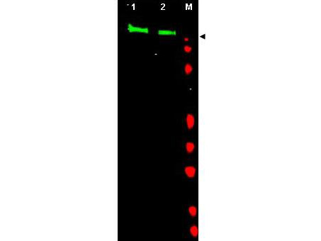
- Western blot using Rockland's Affinity Purified anti-EGFR antibody shows detection of a band at ~170 kDa (lane 1) corresponding to human EGFR present in unstimulated (lane 1) and EGF (50 ng/ml for 15 min) stimulated (lane 2) A431 whole cell lysates (arrowhead). Approximately 30 ug of lysate was separated on a 4-20% Tris-Glycine gel by SDS-PAGE and transferred onto nitrocell-ulose. After blocking the membrane was probed with the primary antibody diluted to 1:1,000. Reaction occurred overnight at 4° C followed by washes and reaction with a 1:10,000 dilution of IRDye800 conjugated Gt-a-Rabbit IgG [H&L] MX (611-132-122) for 45 min at room temperature (800 nm channel, green). Molecular weight estimation was made by comparison to prestained MW markers in lane M (700 nm channel, red). IRDye800 fluorescence image was captured using the Odyssey® Infrared Imaging System developed by LI-COR. IRDye is a trademark of LI-COR, Inc. Other detection systems will yield similar results.
- Validation comment
- Western Blot