 Explore
 Validate
 Learn
Learn Western blot
Antibody data
- Antibody Data
- Antigen structure
- References [1]
- Comments [0]
- Validations
- Western blot [3]
- Immunocytochemistry [1]
- Immunohistochemistry [1]
Submit
Validation data
Reference
Comment
Report error
- Product number
- HPA001336 - Provider product page

- Provider
- Atlas Antibodies
- Proper citation
- Atlas Antibodies Cat#HPA001336, RRID:AB_1078421
- Product name
- Anti-CCDC50
- Antibody type
- Polyclonal
- Reactivity
- Human, Mouse, Rat
- Host
- Rabbit
- Conjugate
- Unconjugated
- Antigen sequence
QEKKDEDIARLLQEKELQEEKKRKKHFPEFPATRA
YADSYYYEDGGMKPRVMKEAVSTPSRMAHRDQEWY
DAEIARKLQEEELLATQVDMRAAQVAQDEEIARLL
MAEEKKAYKKAKEREKSSLDKRKQDPEWKPKTAKA
ANSKSKESDE- Isotype
- IgG
- Vial size
- 100 µl
- Storage
- Store at +4°C for short term storage. Long time storage is recommended at -20°C.
Submitted references Gene knockdown studies revealed CCDC50 as a candidate gene in mantle cell lymphoma and chronic lymphocytic leukemia
Farfsing A, Engel F, Seiffert M, Hartmann E, Ott G, Rosenwald A, Stilgenbauer S, Döhner H, Boutros M, Lichter P, Pscherer A
Leukemia 2009 July;23(11):2018-2026
Leukemia 2009 July;23(11):2018-2026
No comments: Submit comment
Supportive validation
Supportive validation
- Submitted by
- Atlas Antibodies (provider)
- Enhanced method
- Orthogonal validation
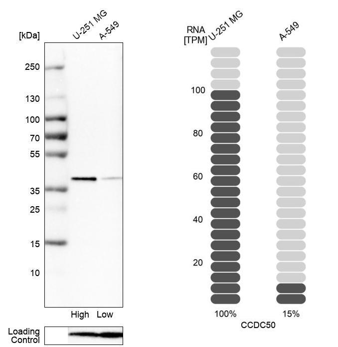
- Main image
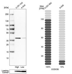
- Experimental details
- Western blot analysis in human cell lines U-251MG and A-549 using Anti-CCDC50 antibody. Corresponding CCDC50 RNA-seq data are presented for the same cell lines. Loading control: Anti-PFN1.
Supportive validation
- Submitted by
- Atlas Antibodies (provider)
- Main image

- Experimental details
- Lane 1: NIH-3T3 cell lysate (Mouse embryonic fibroblast cells)Lane 2: NBT-II cell lysate (Rat Wistar bladder tumour cells)
- Sample type
- MOUSE, RAT

- Submitted by
- Atlas Antibodies (provider)
- Main image

- Experimental details
- Western blot analysis in mouse cell line NIH-3T3 and rat cell line NBT-II.

Supportive validation
- Submitted by
- Atlas Antibodies (provider)
- Main image

- Experimental details
- Immunofluorescent staining of human cell line U-251 MG shows localization to cytosol.
- Sample type
- HUMAN

Supportive validation
- Submitted by
- Atlas Antibodies (provider)
- Main image

- Experimental details
- Immunohistochemical staining of human lymph node shows strong cytoplasmic positivity in a subset of lymphoid cells outside reaction center.
- Sample type
- HUMAN
