 Explore
 Validate
 Learn
LearnPA5-54436
antibody from Invitrogen Antibodies
Targeting: SVEP1
bA427L11.3, C9orf13, FLJ13529, POLYDOM
 Immunohistochemistry
Antibody data
- Antibody Data
- Antigen structure
- References [1]
- Comments [0]
- Validations
- Immunohistochemistry [3]
- Other assay [2]
Submit
Validation data
Reference
Comment
Report error
- Product number
- PA5-54436 - Provider product page

- Provider
- Invitrogen Antibodies
- Product name
- SVEP1 Polyclonal Antibody
- Antibody type
- Polyclonal
- Antigen
- Recombinant full-length protein
- Description
- Immunogen sequence: GDFICTPDNT GVNCTLTCLE GYDFTEGSTD KYYCAYEDGV WKPTYTTEWP DCAKKRFANH GFKSFEMFYK AARCDDTDLM KKFSEAFETT LG
- Concentration
- 0.4 mg/mL
Submitted references SVEP1 as a Genetic Modifier of TEK-Related Primary Congenital Glaucoma.
Young TL, Whisenhunt KN, Jin J, LaMartina SM, Martin SM, Souma T, Limviphuvadh V, Suri F, Souzeau E, Zhang X, Dan Y, Anagnos E, Carmona S, Jody NM, Stangel N, Higuchi EC, Huang SJ, Siggs OM, Simões MJ, Lawson BM, Martin JS, Elahi E, Narooie-Nejad M, Motlagh BF, Quaggin SE, Potter HD, Silva ED, Craig JE, Egas C, Maroofian R, Maurer-Stroh S, Bradfield YS, Tompson SW
Investigative ophthalmology & visual science 2020 Oct 1;61(12):6
Investigative ophthalmology & visual science 2020 Oct 1;61(12):6
No comments: Submit comment
Supportive validation
- Submitted by
- Invitrogen Antibodies (provider)
- Main image
- Experimental details
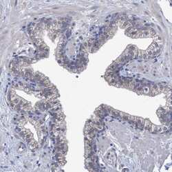
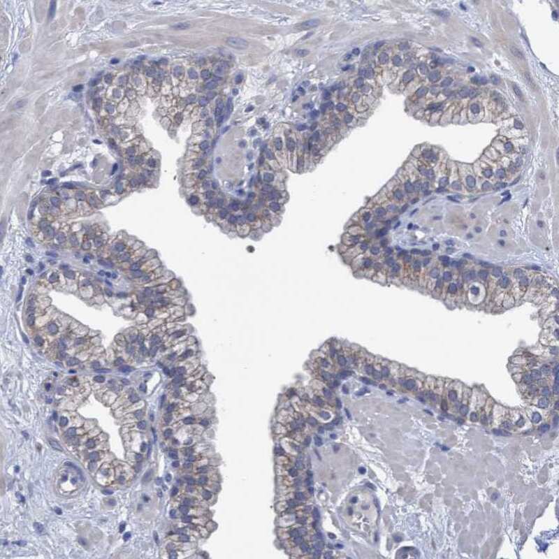
- Immunohistochemical staining of SVEP1 in human prostate using SVEP1 Polyclonal Antibody (Product # PA5-54436) shows weak cytoplasmic positivity in glandular cells.
- Submitted by
- Invitrogen Antibodies (provider)
- Main image
- Experimental details
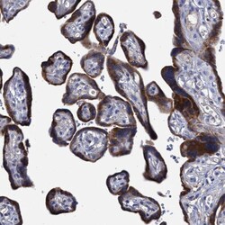
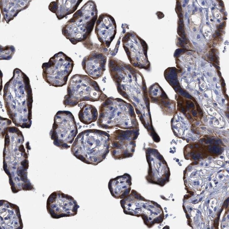
- Immunohistochemical staining of SVEP1 in human placenta using SVEP1 Polyclonal Antibody (Product # PA5-54436) shows strong cytoplasmic positivity in trophoblastic cells.
- Submitted by
- Invitrogen Antibodies (provider)
- Main image
- Experimental details
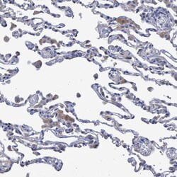
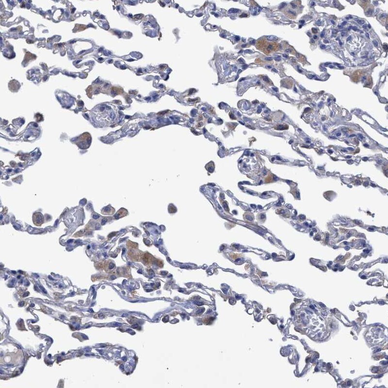
- Immunohistochemical staining of SVEP1 in human lung using SVEP1 Polyclonal Antibody (Product # PA5-54436) shows moderate cytoplasmic positivity in macrophages.
Supportive validation
- Submitted by
- Invitrogen Antibodies (provider)
- Main image
- Experimental details
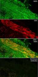
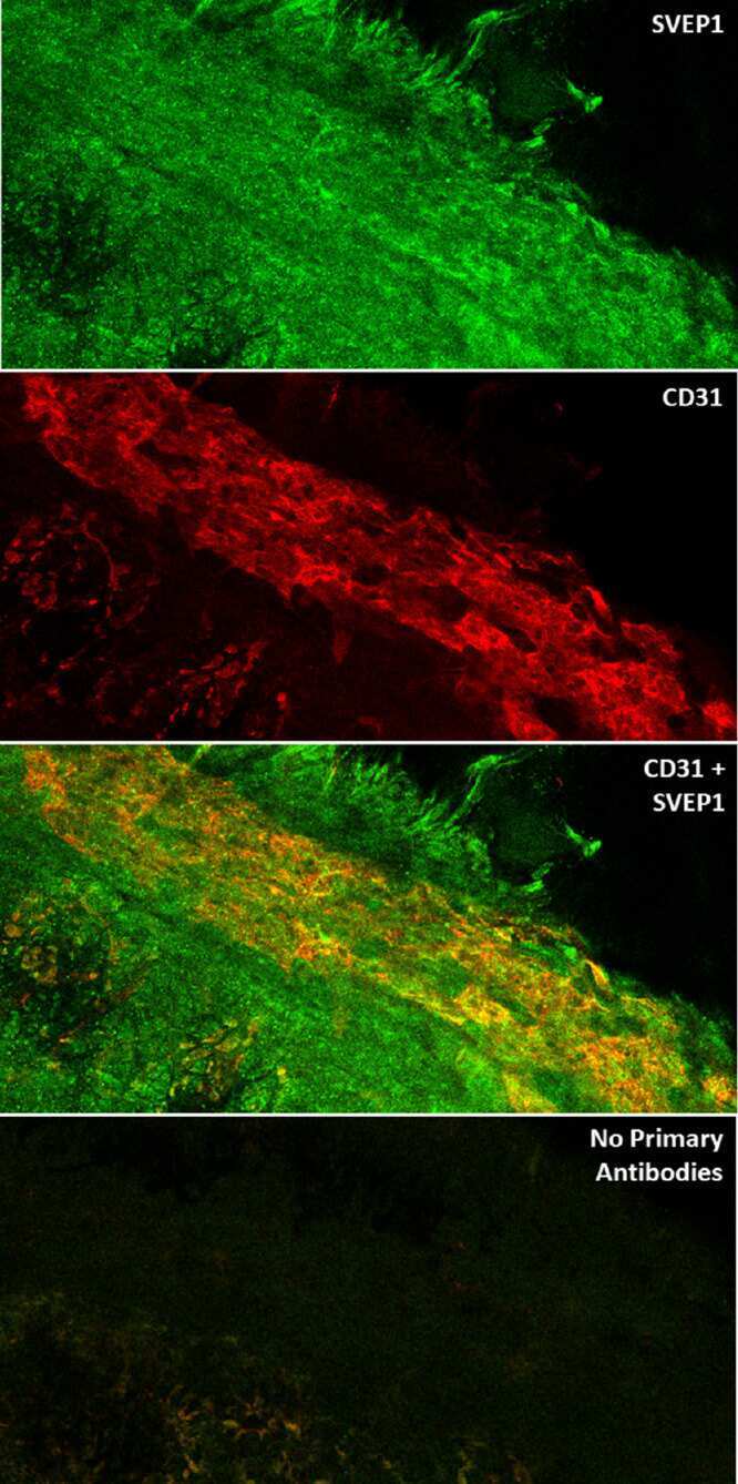
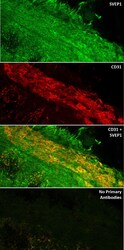
- Figure 6. Immunofluorescent confocal imaging of flat-mounted 1-week old mouse anterior segments stained for SVEP1 and CD31 protein. ( a ) SVEP1 ( green ) expression is observed in the mesenchyme of the developing iridocorneal angle. ( b ) CD31 ( red ), an EC marker, is detected within the interwoven branches of the maturing SC and the ancillary vasculature. ( c ) Overlay revealed mesenchymal-expressed SVEP1 protein in close proximity to the developing SC. ( d ) Anterior segments processed without the addition of SVEP1 and CD31 primary antibodies showed minimal background signal from the secondary antibodies alone.
- Submitted by
- Invitrogen Antibodies (provider)
- Main image
- Experimental details
- Figure 6. Immunofluorescent confocal imaging of flat-mounted 1-week old mouse anterior segments stained for SVEP1 and CD31 protein. ( a ) SVEP1 ( green ) expression is observed in the mesenchyme of the developing iridocorneal angle. ( b ) CD31 ( red ), an EC marker, is detected within the interwoven branches of the maturing SC and the ancillary vasculature. ( c ) Overlay revealed mesenchymal-expressed SVEP1 protein in close proximity to the developing SC. ( d ) Anterior segments processed without the addition of SVEP1 and CD31 primary antibodies showed minimal background signal from the secondary antibodies alone.
