 Explore
 Validate
 Learn
Learn Western blot
 Immunohistochemistry
Antibody data
- Antibody Data
- Antigen structure
- References [0]
- Comments [0]
- Validations
- Immunohistochemistry [2]
- Blocking/Neutralizing [1]
Submit
Validation data
Reference
Comment
Report error
- Product number
- AF1212 - Provider product page

- Provider
- Novus Biologicals
- Product name
- Sheep Polyclonal FGF-16 Antibody
- Antibody type
- Polyclonal
- Description
- Antigen Affinity-purified. Detects human FGF-16 in direct ELISAs and Western blots. In direct ELISAs, less than 1% cross-reactivity with recombinant human (rh) FGF acidic, rhFGF basic, rhFGF-3, -4, -5, -6, -7, -9, -10, -11, -12, -13, -17, -18, -19, -21, -23, recombinant mouse (rm) FGF-8b, and rmFGF-15 is observed.
- Reactivity
- Human, Mouse
- Host
- Sheep
- Conjugate
- Unconjugated
- Isotype
- IgG
- Vial size
- 100 ug
- Concentration
- LYOPH
- Storage
- Use a manual defrost freezer and avoid repeated freeze-thaw cycles. 12 months from date of receipt, -20 to -70 degreesC as supplied. 1 month, 2 to 8 degreesC under sterile conditions after reconstitution. 6 months, -20 to -70 degreesC under sterile conditions after reconstitution.
No comments: Submit comment
Supportive validation
- Submitted by
- Novus Biologicals (provider)
- Main image
- Experimental details
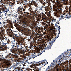
- FGF-16 in Human Heart. FGF-16 was detected in immersion fixed paraffin-embedded sections of human heart using Sheep Anti-Human/Mouse FGF-16 Antigen Affinity-purified Polyclonal Antibody (Catalog # AF1212) at 3 µg/mL overnight at 4 °C. Tissue was stained using the Anti-Sheep HRP-DAB Cell & Tissue Staining Kit (brown; Catalog # CTS019) and counterstained with hematoxylin (blue). Specific staining was localized to cardiomyocytes. View our protocol for Chromogenic IHC Staining of Paraffin-embedded Tissue Sections.

- Submitted by
- Novus Biologicals (provider)
- Main image

- Experimental details
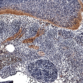
- FGF-16 in Mouse Embryo. FGF-16 was detected in perfusion fixed frozen sections of mouse embryo (13 d.p.c.) using Sheep Anti-Human/Mouse FGF-16 Antigen Affinity-purified Polyclonal Antibody (Catalog # AF1212) at 15 µg/mL overnight at 4 °C. Tissue was stained using the Anti-Sheep HRP-DAB Cell & Tissue Staining Kit (brown; Catalog # CTS019) and counterstained with hematoxylin (blue). Specific staining was localized to roots of dorsal ganglia and spinal cord. View our protocol for Chromogenic IHC Staining of Frozen Tissue Sections.
Supportive validation
- Submitted by
- Novus Biologicals (provider)
- Main image

- Experimental details
- Cell Proliferation Induced by FGF-16 and Neutralization by Human FGF-16 Antibody. Recombinant Human FGF-16 (Catalog # 1212-FG) stimulates proliferation in the NR6R-3T3 mouse fibroblast cell line in a dose-dependent manner (orange line). Proliferation elicited by Recombinant Human FGF-16 (100 ng/mL) is neutralized (green line) by increasing concentrations of Sheep Anti-Human/Mouse FGF-16 Antigen Affinity-purified Polyclonal Antibody (Catalog # AF1212). The ND50 is typically 3-9 µg/mL.
