 Explore
 Validate
 Learn
Learn Western blot
 Immunocytochemistry
Antibody data
- Antibody Data
- Antigen structure
- References [3]
- Comments [0]
- Validations
- Western blot [1]
- Immunohistochemistry [6]
- Flow cytometry [2]
Submit
Validation data
Reference
Comment
Report error
- Product number
- NBP2-01926 - Provider product page

- Provider
- Novus Biologicals
- Product name
- Mouse Monoclonal LIM1 Antibody
- Antibody type
- Monoclonal
- Description
- Affinity purified.
- Reactivity
- Human
- Host
- Mouse
- Isotype
- IgG
- Vial size
- 0.1 ml
- Concentration
- 0.75 mg/ml
- Storage
- Store at -20C. Avoid freeze-thaw cycles.
Submitted references Generating Multiple Kidney Progenitors and Cell Types from Human Pluripotent Stem Cells.
The generation of kidney organoids by differentiation of human pluripotent cells to ureteric bud progenitor-like cells.
Directed differentiation of human pluripotent cells to ureteric bud kidney progenitor-like cells.
Hariharan K, Reinke P, Kurtz A
Methods in molecular biology (Clifton, N.J.) 2019;1926:103-115
Methods in molecular biology (Clifton, N.J.) 2019;1926:103-115
The generation of kidney organoids by differentiation of human pluripotent cells to ureteric bud progenitor-like cells.
Xia Y, Sancho-Martinez I, Nivet E, Rodriguez Esteban C, Campistol JM, Izpisua Belmonte JC
Nature protocols 2014 Nov;9(11):2693-704
Nature protocols 2014 Nov;9(11):2693-704
Directed differentiation of human pluripotent cells to ureteric bud kidney progenitor-like cells.
Xia Y, Nivet E, Sancho-Martinez I, Gallegos T, Suzuki K, Okamura D, Wu MZ, Dubova I, Esteban CR, Montserrat N, Campistol JM, Izpisua Belmonte JC
Nature cell biology 2013 Dec;15(12):1507-15
Nature cell biology 2013 Dec;15(12):1507-15
No comments: Submit comment
Supportive validation
- Submitted by
- Novus Biologicals (provider)
- Main image
- Experimental details
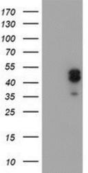
- Western Blot: LIM1 Antibody (2D5) [NBP2-01926] - HEK293T cells were transfected with the pCMV6-ENTRY control (Left lane) or pCMV6-ENTRY LIM1 (Right lane) cDNA for 48 hrs and lysed. Equivalent amounts of cell lysates (5 ug per lane) were separated by SDS-PAGE and immunoblotted with anti-LIM1.

Supportive validation
- Submitted by
- Novus Biologicals (provider)
- Main image

- Experimental details
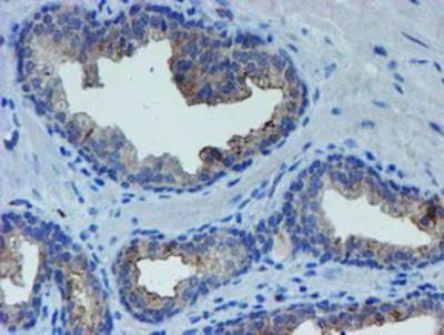
- Immunohistochemistry-Paraffin: LIM1 Antibody (2D5) [NBP2-01926] - Staining of paraffin-embedded Human prostate tissue using anti-LIM1 mouse monoclonal antibody.

- Submitted by
- Novus Biologicals (provider)
- Main image

- Experimental details
- Immunohistochemistry-Paraffin: LIM1 Antibody (2D5) [NBP2-01926] - Staining of paraffin-embedded Human pancreas tissue using anti-LIM1 mouse monoclonal antibody.

- Submitted by
- Novus Biologicals (provider)
- Main image

- Experimental details
- Immunohistochemistry-Paraffin: LIM1 Antibody (2D5) [NBP2-01926] - Staining of paraffin-embedded Human liver tissue using anti-LIM1 mouse monoclonal antibody.

- Submitted by
- Novus Biologicals (provider)
- Main image

- Experimental details
- Immunohistochemistry-Paraffin: LIM1 Antibody (2D5) [NBP2-01926] - Staining of paraffin-embedded Human Kidney tissue using anti-LIM1 mouse monoclonal antibody.

- Submitted by
- Novus Biologicals (provider)
- Main image

- Experimental details
- Immunohistochemistry-Paraffin: LIM1 Antibody (2D5) [NBP2-01926] - Staining of paraffin-embedded Carcinoma of Human prostate tissue using anti-LIM1 mouse monoclonal antibody.
- Submitted by
- Novus Biologicals (provider)
- Main image

- Experimental details
- Immunohistochemistry-Paraffin: LIM1 Antibody (2D5) [NBP2-01926] - Staining of paraffin-embedded Adenocarcinoma of Human endometrium tissue using anti-LIM1 mouse monoclonal antibody.

Supportive validation
- Submitted by
- Novus Biologicals (provider)
- Main image
- Experimental details
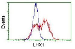
- Flow Cytometry: LIM1 Antibody (2D5) [NBP2-01926] - HEK293T cells transfected with either overexpression plasmid (Red) or empty vector control plasmid (Blue) were immunostained by anti-LIM1 antibody, and then analyzed by flow cytometry.

- Submitted by
- Novus Biologicals (provider)
- Main image
- Experimental details
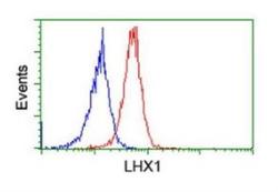
- Flow Cytometry: LIM1 Antibody (OTI2D5) [NBP2-01926] - Analysis of Jurkat cells, using anti-LHX1 antibody (Red) compared to a nonspecific negative control antibody (Blue).
