 Explore
 Validate
 Learn
LearnPA5-28749
antibody from Invitrogen Antibodies
Targeting: ESRRA
ERR1, ERRa, ERRalpha, ESRL1, NR3B1
 Western blot Immunocytochemistry
 Immunoprecipitation Immunohistochemistry Chromatin Immunoprecipitation Other assay
Antibody data
- Antibody Data
- Antigen structure
- References [0]
- Comments [0]
- Validations
- Western blot [7]
- Immunocytochemistry [2]
- Immunohistochemistry [3]
- Chromatin Immunoprecipitation [2]
- Other assay [1]
Submit
Validation data
Reference
Comment
Report error
- Product number
- PA5-28749 - Provider product page

- Provider
- Invitrogen Antibodies
- Product name
- ESRRA Polyclonal Antibody
- Antibody type
- Polyclonal
- Antigen
- Synthetic peptide
- Reactivity
- Human, Mouse, Rat
- Host
- Rabbit
- Isotype
- IgG
- Vial size
- 100 µL
- Concentration
- 1.32 mg/mL
- Storage
- Store at 4°C short term. For long term storage, store at -20°C, avoiding freeze/thaw cycles.
No comments: Submit comment
Supportive validation
- Submitted by
- Invitrogen Antibodies (provider)
- Main image
- Experimental details
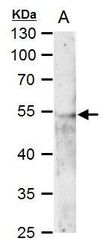
- Western blot analysis of ERR alpha using 50 µg mouse kideny extract. Samples were loaded onto a 10% SDS-PAGE gel and probed with an ERR alpha polyclonal antibody (Product # PA5-28749) at a dilution of 1:500.

- Submitted by
- Invitrogen Antibodies (provider)
- Main image

- Experimental details
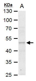
- Western blot analysis of ERR alpha using 50 µg rat kideny extract. Samples were loaded onto a 10% SDS-PAGE gel and probed with an ERR alpha polyclonal antibody (Product # PA5-28749) at a dilution of 1:500.
- Submitted by
- Invitrogen Antibodies (provider)
- Main image

- Experimental details
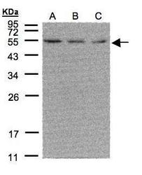
- Western Blot using ESRRA Polyclonal Antibody (Product # PA5-28749). Sample (30 µg of whole cell lysate). A: 293T. B: A431. C: H1299. 12% SDS PAGE. ESRRA Polyclonal Antibody (Product # PA5-28749) diluted at 1:500. The HRP-conjugated anti-rabbit IgG antibody was used to detect the primary antibody.

- Submitted by
- Invitrogen Antibodies (provider)
- Main image
- Experimental details
- Western Blot using ESRRA Polyclonal Antibody (Product # PA5-28749). Sample (30 µg of whole cell lysate). A: 293T. B: A431. C: H1299. 12% SDS PAGE. ESRRA Polyclonal Antibody (Product # PA5-28749) diluted at 1:500. The HRP-conjugated anti-rabbit IgG antibody was used to detect the primary antibody.
- Submitted by
- Invitrogen Antibodies (provider)
- Main image

- Experimental details
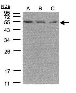
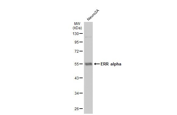
- Western Blot using ESRRA Polyclonal Antibody (Product # PA5-28749). Whole cell extract (30 µg) was separated by 10% SDS-PAGE, and the membrane was blotted with ESRRA Polyclonal Antibody (Product # PA5-28749) diluted at 1:500. The HRP-conjugated anti-rabbit IgG antibody was used to detect the primary antibody.
- Submitted by
- Invitrogen Antibodies (provider)
- Main image
- Experimental details
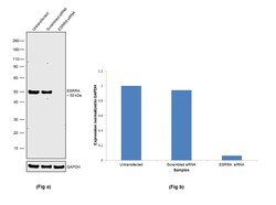
- Knockdown of ESRRA was achieved by transfecting SK-BR-3 with ESRRA specific siRNAs (Silencer® select Product # s4831). Western blot analysis (Fig. a) was performed using whole cell extracts from the ESRRA knockdown cells (lane 3), non-specific scrambled siRNA transfected cells (lane 2) and untransfected cells (lane 1). The blot was probed with ESRRA Polyclonal Antibody (Product # PA5-28749, 1:1000 dilution) and Goat anti-Rabbit IgG (H+L) Superclonal™ Secondary Antibody, HRP (Product # A27036, 1:4000 dilution). Densitometric analysis of this western blot is shown in histogram (Fig. b). Decrease in signal upon siRNA mediated knock down confirms that antibody is specific to ESRRA.

- Submitted by
- Invitrogen Antibodies (provider)
- Main image
- Experimental details
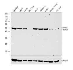
- Western blot was performed using Anti-ESRRA Polyclonal Antibody (Product # PA5-28749) and a 50 kDa band corresponding to ESRRA was observed across the cell lines and tissues tested except in MCF 10A which is reported to be negative. Modified Whole cell extracts (1% SDS) (30 µg lysate) of SK-BR-3 (Lane 1), MCF7 (Lane 2), T-47D (Lane 3), MCF 10A (Lane 4), PC-3 (Lane 5), OVCAR-3 (Lane 6), A-431 (Lane 7), Mouse Kidney (Lane 8) and Rat Liver (Lane 9) were electrophoresed using NuPAGE™ 10% Bis-Tris Protein Gel (Product # NP0302BOX). Resolved proteins were then transferred onto a nitrocellulose membrane (Product # IB23001) by iBlot® 2 Dry Blotting System (Product # IB21001). The blot was probed with the primary antibody (1:1000 dilution) and detected by chemiluminescence with Goat anti-Rabbit IgG (H+L), Superclonal™ Recombinant Secondary Antibody, HRP (Product # A27036, 1:4000 dilution) using the iBright FL 1000 (Product # A32752). Chemiluminescent detection was performed using Novex® ECL Chemiluminescent Substrate Reagent Kit (Product # WP20005).

Supportive validation
- Submitted by
- Invitrogen Antibodies (provider)
- Main image
- Experimental details
- Immunocytochemistry analysis of ESRRA in MCF-7 cells. Samples were incubated in ESRRA polyclonal antibody (Product # PA5-28749) using a dilution of 1:500. Sample: MCF-7 cells were fixed in 4% paraformaldehyde at RT for 15 min. Green: ERR alpha stained by ERR alpha antibody [N1], N-term. Red: alpha Tubulin, a cytoskeleton marker, stained by alpha Tubulin antibody [GT114] diluted at 1:1,000.

- Submitted by
- Invitrogen Antibodies (provider)
- Main image

- Experimental details
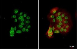
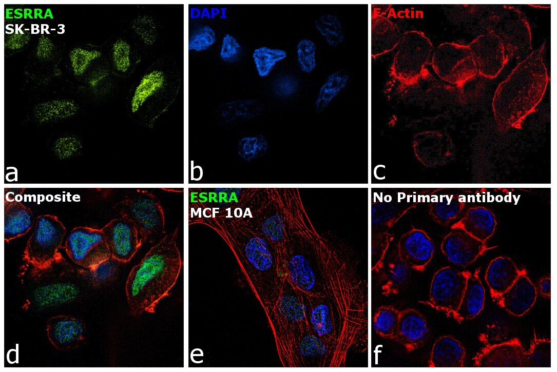
- Immunofluorescence analysis of ESRRA was performed using 70% confluent log phase SK-BR-3 and MCF 10A cells. The cells were fixed with 4% paraformaldehyde for 10 minutes, permeabilized with 0.1% Triton™ X-100 for 15 minutes, and blocked with 2% BSA for 1 hour at room temperature. The cells were labeled with ESRRA Polyclonal Antibody (Product # PA5-28749) at 1:100 dilution in 0.1% BSA, incubated at 4 degree celsius overnight and then with Goat anti-Rabbit IgG (H+L), Superclonal™ Recombinant Secondary Antibody, Alexa Fluor 488 conjugate (Product # A27034) at a dilution of 1:2000 for 45 minutes at room temperature (Panel a: green). Nuclei (Panel b: blue) were stained with SlowFade® Gold Antifade Mountant with DAPI (Product # S36938). F-actin (Panel c: red) was stained with Rhodamine Phalloidin (Product # R415, 1:300). Panel d represents the merged image showing predominantly nuclear localization. Panel e represents MCF 10A cells having low expression of ESRRA. Panel f represents control cells with no primary antibody to assess background. The images were captured at 60X magnification.
Supportive validation
- Submitted by
- Invitrogen Antibodies (provider)
- Main image

- Experimental details
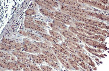
- Immunohistochemistry analysis of ESRRA in paraffin-embedded mouse stomach. Samples were incubated in ESRRA polyclonal antibody (Product # PA5-28749) using a dilution of 1:500. Antigen Retrieval: Citrate buffer, pH 6.0, 15 min.
- Submitted by
- Invitrogen Antibodies (provider)
- Main image
- Experimental details
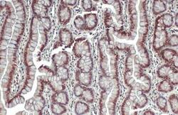
- Immunohistochemistry analysis of ESRRA in paraffin-embedded rat colon. Samples were incubated in ESRRA polyclonal antibody (Product # PA5-28749) using a dilution of 1:500. Antigen Retrieval: Citrate buffer, pH 6.0, 15 min.

- Submitted by
- Invitrogen Antibodies (provider)
- Main image

- Experimental details
- Immunohistochemistry analysis of ESRRA in paraffin-embedded mouse intestine. Samples were incubated in ESRRA polyclonal antibody (Product # PA5-28749) using a dilution of 1:500. Antigen Retrieval: Citrate buffer, pH 6.0, 15 min.

Supportive validation
- Submitted by
- Invitrogen Antibodies (provider)
- Main image
- Experimental details
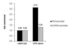
- Cross-linked ChIP was performed with MCF-7 chromatin extract and 5 µg of either control rabbit IgG or ESRRA Polyclonal Antibody (Product # PA5-28749). The precipitated DNA was detected by PCR with primer set targeting to PS2 promoter or GAPDH promoter.

- Submitted by
- Invitrogen Antibodies (provider)
- Main image

- Experimental details
- Chromatin Immunoprecipitation (ChIP) assay of endogenous ESRRA protein using Anti-ESRRA Antibody: ChIP was performed using Anti-ESRRA Rabbit Polyclonal Antibody (Product # PA5-28749) 5 µg, on sheared chromatin from etoposide treated MCF-7 cells using the MAGnify ChIP System kit (Product # 49-2024). Normal Rabbit IgG was used as a negative IP control. The purified DNA was analyzed by qPCR using primers binding to promoter of OGDH, IDH3A promoter and SAT2 satellite repeats. Data is presented as fold enrichment of the antibody signal versus the negative control IgG using the comparative CT method.

Supportive validation
- Submitted by
- Invitrogen Antibodies (provider)
- Main image

- Experimental details
- ERR alpha antibody immunoprecipitates ERR alpha protein in IP experiments. IP Sample: 293T whole cell lysate/extract A. 40 µg 293T whole cell lysate/extract B. Control with 2 µg of preimmune rabbit IgG C. Immunoprecipitation of ERR alpha protein by 2 µg of ERR alpha antibody (Product # PA5-28749) 7.5% SDS-PAGE The immunoprecipitated ERR alpha protein was detected by ERR alpha antibody (Product # PA5-28749) diluted at 1:1,000.
