 Explore
 Validate
 Learn
LearnPA5-36391
antibody from Invitrogen Antibodies
Targeting: NCOA4
ARA70, DKFZp762E1112, ELE1, PTC3, RFG
 Western blot
Antibody data
- Antibody Data
- Antigen structure
- References [1]
- Comments [0]
- Validations
- Western blot [1]
- Immunohistochemistry [2]
- Other assay [2]
Submit
Validation data
Reference
Comment
Report error
- Product number
- PA5-36391 - Provider product page

- Provider
- Invitrogen Antibodies
- Product name
- NCOA4 Polyclonal Antibody
- Antibody type
- Polyclonal
- Antigen
- Synthetic peptide
- Description
- This antibody detects endogenous protein at a molecular weight of 70 kDa. Purity is >95% by SDS-PAGE.
- Reactivity
- Human, Mouse, Rat
- Host
- Rabbit
- Isotype
- IgG
- Vial size
- 100 µL
- Concentration
- 1 mg/mL
- Storage
- Store at 4°C short term. For long term storage, store at -20°C, avoiding freeze/thaw cycles.
Submitted references Involvement of oxidative stress-induced annulus fibrosus cell and nucleus pulposus cell ferroptosis in intervertebral disc degeneration pathogenesis.
Yang RZ, Xu WN, Zheng HL, Zheng XF, Li B, Jiang LS, Jiang SD
Journal of cellular physiology 2021 Apr;236(4):2725-2739
Journal of cellular physiology 2021 Apr;236(4):2725-2739
No comments: Submit comment
Supportive validation
- Submitted by
- Invitrogen Antibodies (provider)
- Main image

- Experimental details
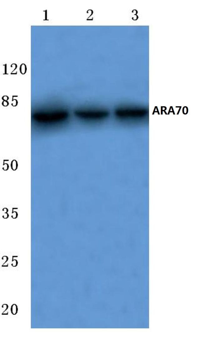
- Western blot analysis of ARA70 using ARA70 polyclonal antibody (Product # PA5-36391) at a dilution of 1:500. Lane 1: MCF-7 cell lysate, Lane 2: Mouse liver tissue lysate, Lane 3: Rat lung tissue lysate.
Supportive validation
- Submitted by
- Invitrogen Antibodies (provider)
- Main image
- Experimental details
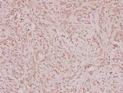
- Immunohistochemical analysis of ARA70 in paraffin-embedded human breast carcinoma using ARA70 polyclonal antibody (Product # PA5-36391) at a dilution of 1:100.

- Submitted by
- Invitrogen Antibodies (provider)
- Main image
- Experimental details
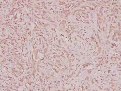
- Immunohistochemistry analysis of NCOA4 in paraffin-embedded human breast carcinoma tissue. Samples were incubated with NCOA4 polyclonal antibody (Product # PA5-36391) at a dilution of 1:100.

Supportive validation
- Submitted by
- Invitrogen Antibodies (provider)
- Main image

- Experimental details
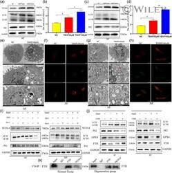
- 3 Figure Cell death of annulus fibrosus cells (AFCs) and nucleus pulposus cells (NPCs) induced by tert -butyl hydroperoxide (TBHP) was inhibited and was similar to RSL3-induced AFCs and NPCs ferroptosis. AFCs and NPCs were treated with 100 muM TBHP with or without the specific inhibitors deferoxamine mesylate (DFO; 10 muM) or ferrostatin-1 (Fer-1; 5 muM) or were exposed to RSL3 at different concentration (0, 10, 15, 20 muM). (a) Western blotting assay of nuclear receptor coactivator 4 (NCOA4), glutathione peroxidase 4 (GPX4), ferritin heavy chain (FTH), and GAPDH in AFCs. (b,c) The difference in GPX4, NCOA4, and FTH expression in AFCs under different treatments. (d) Lipid peroxidation level under different treatment conditions of AFCs. (e) Immunofluorescence detection of FTH in AFCs. (f) Western blotting assay of NCOA4, GPX4, FTH, and GAPDH in NPCs. (g,h) The difference in GPX4, NCOA4, and FTH expression in NPCs under different treatments. (i) Lipid peroxidation level under different treatment conditions of NPCs. (j) Immunofluorescence detection of FTH in NPCs. *** p < .001, ** p < .01, * p < .05 ( n = 3)

- Submitted by
- Invitrogen Antibodies (provider)
- Main image
- Experimental details
- 4 Figure tert -Butyl hydroperoxide (TBHP)-induced ferroptosis of annulus fibrosus cells (AFCs) and nucleus pulposus cells (NPCs) in an autophagy-dependent manner. (a) Protein expression of nuclear receptor coactivator 4 (NCOA4), LC3, P62, and GAPDH in AFCs exposed to TBHP. (b) The ratio of LC3II/LC3I in AFCs under different concentrations of TBHP. (c) Protein expression of NCOA4, LC3, P62, and GAPDH in NPCs exposed to TBHP. (d) The ratio of LC3II/LC3I in NPCs under different concentrations of TBHP. (e) Transmission electron microscopy showed increased autophagosomes of AFCs after TBHP stimulation. (f) Fluorescence images of AFCs transfected with mRFP-GFP-LC3 adenovirus. The promotion of Red dot represents that the autophagy was enhanced. (g) Transmission electron microscopy showed increased autophagosomes of NPCs after TBHP stimulation. (h) Fluorescence images of NPCs infected with mRFP-GFP-LC3 adenovirus showed the level of autophagy promoted after TBHP treatment. (i) AFCs and NPCs were exposed to 100 muM TBHP with or without deferoxamine mesylate (DFO; 10 muM) or ferrostatin-1 (Fer-1; 5 muM) for 3 h. The cell lysates were analyzed for the expression of NCOA4, LC3, P62, GAPDH by western blotting. (j) AFCs and NPCs were treated with TBHP (100 muM) with or without 3-methyladenine (3-MA; 10 muM). Western blotting assay of LC3, P62, glutathione peroxidase 4 (GPX4), ferritin heavy chain (FTH), and GAPDH in AFCs and NPCs. (k) Co-immunoprecipitation (Co-IP) test analyzed the relati
