Explore
 Validate
 Learn
Learn Western blot
Antibody data
- Antibody Data
- Antigen structure
- References [0]
- Comments [0]
- Validations
- Western blot [1]
- Immunocytochemistry [1]
- Immunohistochemistry [2]
Submit
Validation data
Reference
Comment
Report error
- Product number
- MA5-31334 - Provider product page

- Provider
- Invitrogen Antibodies
- Product name
- STX7 Monoclonal Antibody (CL0257)
- Antibody type
- Monoclonal
- Antigen
- Recombinant full-length protein
- Description
- Immunogen sequence: GVGGDPAQLA QRISSNIQKI TQCSVEIQRT LNQLGTPQDS PELRQQLQQK QQYTNQLAKE TDKYIKEFGS LPTTPSEQRQ RKIQKDRLVA EFTTSLTNFQ KVQRQAAERE KEFVARVRAS SRVSGSFPED SSKERNLVSW ESQTQPQV
- Reactivity
- Human
- Host
- Mouse
- Isotype
- IgG
- Antibody clone number
- CL0257
- Vial size
- 100 µL
- Concentration
- 1 mg/mL
- Storage
- Store at 4°C short term. For long term storage, store at -20°C, avoiding freeze/thaw cycles.
No comments: Submit comment
Supportive validation
- Submitted by
- Invitrogen Antibodies (provider)
- Main image

- Experimental details
- Western blot analysis of STX7 by a STX7 monoclonal antibody (Product # MA5-31334). Lane 1: Marker [kDa] Lane 2: Human tonsil tissue lysate.

Supportive validation
- Submitted by
- Invitrogen Antibodies (provider)
- Main image

- Experimental details
- Immunocytochemistry-Immunofluorescence analysis of STX7 in U-251 cells using STX7 Monoclonal Antibody (CL0257) (Product # MA5-31334), showing specific staining in vesicles in green. Microtubule- and nuclear probes are visualized in red and blue, respectively (where available).

Supportive validation
- Submitted by
- Invitrogen Antibodies (provider)
- Main image

- Experimental details
- Immunohistochemical analysis of STX7 in human melanoma using a STX7 monoclonal antibody (Product # MA5-31334). The analysis shows strong cytoplasmic and membrane positivity in tumour cells.

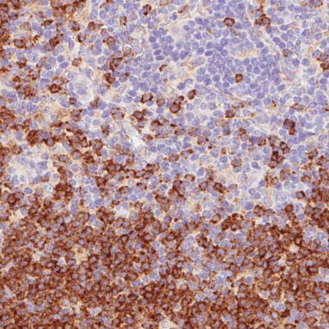
- Submitted by
- Invitrogen Antibodies (provider)
- Main image

- Experimental details
- Immunohistochemical analysis of STX7 in human lymph node using a STX7 monoclonal antibody (Product # MA5-31334). The analysis shows strong membrane positivity in non-germinal center cells.