Explore
 Validate
 Learn
LearnMAB25341
antibody from Novus Biologicals
Targeting: MRC1
bA541I19.1, CD206, CLEC13D, CLEC13DL, MRC1L1
 Western blot
Antibody data
- Antibody Data
- Antigen structure
- References [3]
- Comments [0]
- Validations
- Western blot [2]
- Immunohistochemistry [1]
Submit
Validation data
Reference
Comment
Report error
- Product number
- MAB25341 - Provider product page

- Provider
- Novus Biologicals
- Product name
- Mouse Monoclonal MMR/CD206/Mannose Receptor Antibody
- Antibody type
- Monoclonal
- Description
- Protein A or G purified from hybridoma culture supernatant. Detects human MMR/CD206 in direct ELISAs and Western blots. In direct ELISAs, no cross-reactivity with recombinant mouse (rm) MMR or recombinant human Mrc2 is observed. In Western blots, approximately 25% cross-reactivity with rmMMR is observed.
- Reactivity
- Human
- Host
- Mouse
- Isotype
- IgG
- Vial size
- 100 ug
- Concentration
- LYOPH
- Storage
- Use a manual defrost freezer and avoid repeated freeze-thaw cycles. 12 months from date of receipt, -20 to -70 degreesC as supplied. 1 month, 2 to 8 degreesC under sterile conditions after reconstitution. 6 months, -20 to -70 degreesC under sterile conditions after reconstitution.
Submitted references Macrophage Mannose Receptor CD206 Predicts Prognosis in Community-acquired Pneumonia.
Regional Differences Between Perisynovial and Infrapatellar Adipose Tissue Depots and Their Response to Class II and Class III Obesity in Patients With Osteoarthritis.
Macrophage Activation in Pediatric Nonalcoholic Fatty Liver Disease (NAFLD) Correlates with Hepatic Progenitor Cell Response via Wnt3a Pathway.
Tsuchiya K, Suzuki Y, Yoshimura K, Yasui H, Karayama M, Hozumi H, Furuhashi K, Enomoto N, Fujisawa T, Nakamura Y, Inui N, Yokomura K, Suda T
Scientific reports 2019 Dec 10;9(1):18750
Scientific reports 2019 Dec 10;9(1):18750
Regional Differences Between Perisynovial and Infrapatellar Adipose Tissue Depots and Their Response to Class II and Class III Obesity in Patients With Osteoarthritis.
Harasymowicz NS, Clement ND, Azfer A, Burnett R, Salter DM, Simpson AHWR
Arthritis & rheumatology (Hoboken, N.J.) 2017 Jul;69(7):1396-1406
Arthritis & rheumatology (Hoboken, N.J.) 2017 Jul;69(7):1396-1406
Macrophage Activation in Pediatric Nonalcoholic Fatty Liver Disease (NAFLD) Correlates with Hepatic Progenitor Cell Response via Wnt3a Pathway.
Carpino G, Nobili V, Renzi A, De Stefanis C, Stronati L, Franchitto A, Alisi A, Onori P, De Vito R, Alpini G, Gaudio E
PloS one 2016;11(6):e0157246
PloS one 2016;11(6):e0157246
No comments: Submit comment
Supportive validation
- Submitted by
- Novus Biologicals (provider)
- Main image

- Experimental details
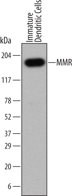
- Detection of Human MMR/CD206 by Western Blot. Western blot shows lysates of human immature dendritic cells. PVDF Membrane was probed with 1 µg/mL of Mouse Anti-Human MMR/CD206 Monoclonal Antibody (Catalog # MAB25341) followed by HRP-conjugated Anti-Mouse IgG Secondary Antibody (Catalog # HAF007). A specific band was detected for MMR/CD206 at approximately 150 kDa (as indicated). This experiment was conducted under reducing conditions and using Immunoblot Buffer Group 1.
- Submitted by
- Novus Biologicals (provider)
- Main image

- Experimental details
- Detection of Human MMR/CD206 by Simple WesternTM. Simple Western lane view shows lysates of human immature dendritic cells, loaded at 0.2 mg/mL. A specific band was detected for MMR/CD206 at approximately 228 kDa (as indicated) using 20 µg/mL of Mouse Anti-Human MMR/CD206 Monoclonal Antibody (Catalog # MAB25341). This experiment was conducted under reducing conditions and using the 66-440 kDa separation system.

Supportive validation
- Submitted by
- Novus Biologicals (provider)
- Main image

- Experimental details
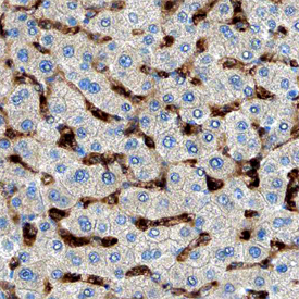
- MMR/CD206 in Human Liver. MMR/CD206 was detected in immersion fixed paraffin-embedded sections of human liver using Mouse Anti-Human MMR/CD206 Monoclonal Antibody (Catalog # MAB25341) at 15 µg/mL overnight at 4 °C. Before incubation with the primary antibody, tissue was subjected to heat-induced epitope retrieval using Antigen Retrieval Reagent-Basic (Catalog # CTS013). Tissue was stained using the Anti-Mouse HRP-DAB Cell & Tissue Staining Kit (brown; Catalog # CTS002) and counterstained with hematoxylin (blue). Specific staining was localized to endothelial cells in sinusoids. View our protocol for Chromogenic IHC Staining of Paraffin-embedded Tissue Sections.