 Explore
 Validate
 Learn
Learn710001
antibody from Invitrogen Antibodies
Targeting: CCL5
D17S136E, MGC17164, RANTES, SCYA5, SISd, TCP228
 Western blot
Antibody data
- Antibody Data
- Antigen structure
- References [4]
- Comments [0]
- Validations
- Western blot [1]
- Immunocytochemistry [1]
- Immunohistochemistry [1]
- Other assay [1]
Submit
Validation data
Reference
Comment
Report error
- Product number
- 710001 - Provider product page

- Provider
- Invitrogen Antibodies
- Product name
- CCL5 (RANTES) Recombinant Polyclonal Antibody (25HCLC)
- Antibody type
- Polyclonal
- Antigen
- Other
- Description
- This antibody is predicted to react with rat and human based on sequence homology.
- Antibody clone number
- 25HCLC
- Concentration
- 0.5 mg/mL
Submitted references PARP inhibitors promote stromal fibroblast activation by enhancing CCL5 autocrine signaling in ovarian cancer.
Dysregulated oxalate metabolism is a driver and therapeutic target in atherosclerosis.
DSE regulates the malignant characters of hepatocellular carcinoma cells by modulating CCL5/CCR1 axis.
NADPH Oxidase 4 Regulates Inflammation in Ischemic Heart Failure: Role of Soluble Epoxide Hydrolase.
Li X, Fang T, Xu S, Jin P, Zhou D, Wang Z, Li H, Yang Z, Chen G, Zheng X, Xia Y, Wei X, Zhang Z, Yang X, Wang Y, Gao Q
NPJ precision oncology 2021 Jun 9;5(1):49
NPJ precision oncology 2021 Jun 9;5(1):49
Dysregulated oxalate metabolism is a driver and therapeutic target in atherosclerosis.
Liu Y, Zhao Y, Shukha Y, Lu H, Wang L, Liu Z, Liu C, Zhao Y, Wang H, Zhao G, Liang W, Fan Y, Chang L, Yurdagul A Jr, Pattillo CB, Orr AW, Aviram M, Wen B, Garcia-Barrio MT, Zhang J, Liu W, Sun D, Hayek T, Chen YE, Rom O
Cell reports 2021 Jul 27;36(4):109420
Cell reports 2021 Jul 27;36(4):109420
DSE regulates the malignant characters of hepatocellular carcinoma cells by modulating CCL5/CCR1 axis.
Liao WC, Yen HR, Liao CK, Tseng TJ, Lan CT, Liu CH
American journal of cancer research 2019;9(2):347-362
American journal of cancer research 2019;9(2):347-362
NADPH Oxidase 4 Regulates Inflammation in Ischemic Heart Failure: Role of Soluble Epoxide Hydrolase.
Stevenson MD, Canugovi C, Vendrov AE, Hayami T, Bowles DE, Krause KH, Madamanchi NR, Runge MS
Antioxidants & redox signaling 2019 Jul 1;31(1):39-58
Antioxidants & redox signaling 2019 Jul 1;31(1):39-58
No comments: Submit comment
Supportive validation
- Submitted by
- Invitrogen Antibodies (provider)
- Main image

- Experimental details
- Western blot analysis of recombinant mouse RANTES (5 ng, lane1) and recombinant rat Rantes (5 ng, lane 2) using a RANTES Recombinant Rabbit Polyclonal Antibody (Product # 710001) at a dilution of 0.1 µg/mL. NBT/BCIP was used as the substrate (Product # WB7105).

Supportive validation
- Submitted by
- Invitrogen Antibodies (provider)
- Main image
- Experimental details
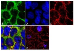
- Immunofluorescence analysis of CCL5/RANTES was performed using 70% confluent log phase HEK-293 cells. The cells were fixed with 4% paraformaldehyde for 10 minutes, permeabilized with 0.1% Triton™ X-100 for 10 minutes, and blocked with 2% BSA for 1 hour at room temperature. The cells were labeled with CCL5/RANTES (25HCLC) Recombinant Rabbit Polyclonal Antibody (Product # 710001) at 2 µg/mL in 0.1% BSA and incubated for 3 hours at room temperature and then labeled with Goat anti-Rabbit IgG (H+L) Superclonal™ Secondary Antibody, Alexa Fluor® 488 conjugate (Product # A27034) a dilution of 1:2000 for 45 minutes at room temperature (Panel a: green). Nuclei (Panel b: blue) were stained with SlowFade® Gold Antifade Mountant with DAPI (Product # S36938). F-actin (Panel c: red) was stained with Alexa Fluor® 555 Rhodamine Phalloidin (Product # R415, 1:300). Panel d represents the merged image showing cytoplasmic localization. Panel e shows the no primary antibody control. The images were captured at 60X magnification.

Supportive validation
- Submitted by
- Invitrogen Antibodies (provider)
- Main image

- Experimental details
- Immunohistochemistry analysis of CCL5/RANTES showing staining in the cytoplasm of paraffin-embedded mouse lung tissue (right) compared to a negative control without primary antibody (left). To expose target proteins, antigen retrieval was performed using 10mM sodium citrate (pH 6.0), microwaved for 8-15 min. Following antigen retrieval, tissues were blocked in 3% H2O2-methanol for 15 min at room temperature, washed with ddH2O and PBS, and then probed with a CCL5/RANTES Recombinant Rabbit Polyclonal Antibody (Product # 710001) diluted in 3% BSA-PBS at a dilution of 1:20 for 1 hour at 37ºC in a humidified chamber. Tissues were washed extensively in PBST and detection was performed using an HRP-conjugated secondary antibody followed by colorimetric detection using a DAB kit. Tissues were counterstained with hematoxylin and dehydrated with ethanol and xylene to prep for mounting.

Supportive validation
- Submitted by
- Invitrogen Antibodies (provider)
- Main image

- Experimental details
- Fig. 7 Olaparib induces stromal fibroblast activation in human OC tumor specimens. a Immunohistochemical detection of alpha-SMA in matched tumor specimens from patients with OC before and after administration with Ola. Scale bar, 50 um. b Quantification of alpha-SMA-positive areas in tumor specimens from OC patients. Same images were measured repeatedly. c - e Representative images and quantification of Masson's trichrome and picrosirius red staining showing that Ola administration increased the content of stromal components and collagen deposition in human OC tumor specimens. Scale bar, 50 um. Same images were measured repeatedly. f , g Representative IHC images and quantification of CCL5 and p-P65 in matched tumor specimens from patients with OC before and after administration with Ola. Scale bar, 50 um. Measures were taken from different samples ( n = 6). h Correlation analysis of CCL5 IHC scores and alpha-SMA IHC scores in matched tumor specimens from patients with OC before and after administration with Ola. i Correlation analysis of p-P65 IHC scores and CCL5 IHC scores in matched tumor specimens from patients with OC before and after administration with Ola. j Schematic representation of the effect of PARPis on human ovarian stromal fibroblasts. Data are expressed as mean +- s.e.m., * p < 0.05; ** p < 0.01; *** p < 0.001.
