 Explore
 Validate
 Learn
Learn Western blot
 ELISA
ELISAAntibody data
- Antibody Data
- Antigen structure
- References [17]
- Comments [0]
- Validations
- Western blot [2]
- Immunohistochemistry [7]
- Other assay [5]
Submit
Validation data
Reference
Comment
Report error
- Product number
- 13-0400 - Provider product page

- Provider
- Invitrogen Antibodies
- Product name
- NEFL Monoclonal Antibody (DA2)
- Antibody type
- Monoclonal
- Antigen
- Other
- Description
- 13-0400 detects the neurofilament, light chain in human, rat, and mouse samples.
- Reactivity
- Human, Mouse, Rat
- Host
- Mouse
- Isotype
- IgG
- Antibody clone number
- DA2
- Vial size
- 200 µg
- Concentration
- 0.5 mg/mL
- Storage
- -20°C
Submitted references Progenitor cells from brown adipose tissue undergo neurogenic differentiation.
Cep215 is essential for morphological differentiation of astrocytes.
Impaired Neurofilament Integrity and Neuronal Morphology in Different Models of Focal Cerebral Ischemia and Human Stroke Tissue.
Unbiased Proteomics of Early Lewy Body Formation Model Implicates Active Microtubule Affinity-Regulating Kinases (MARKs) in Synucleinopathies.
Substituting mouse transcription factor Pou4f2 with a sea urchin orthologue restores retinal ganglion cell development.
Muse Cells, a New Type of Pluripotent Stem Cell Derived from Human Fibroblasts.
Repeated Three-Hour Maternal Separation Induces Depression-Like Behavior and Affects the Expression of Hippocampal Plasticity-Related Proteins in C57BL/6N Mice.
Ginseng Total Saponins Reverse Corticosterone-Induced Changes in Depression-Like Behavior and Hippocampal Plasticity-Related Proteins by Interfering with GSK-3 β -CREB Signaling Pathway.
Phagocytosis of neuronal debris by microglia is associated with neuronal damage in multiple sclerosis.
Beneficial effects of benzodiazepine diazepam on chronic stress-induced impairment of hippocampal structural plasticity and depression-like behavior in mice.
Near complete loss of retinal ganglion cells in the math5/brn3b double knockout elicits severe reductions of other cell types during retinal development.
Changes in NMDA receptor subunits and interacting PSD proteins in dorsolateral prefrontal and anterior cingulate cortex indicate abnormal regional expression in schizophrenia.
A NUDEL-dependent mechanism of neurofilament assembly regulates the integrity of CNS neurons.
The TSC1 tumor suppressor hamartin interacts with neurofilament-L and possibly functions as a novel integrator of the neuronal cytoskeleton.
Brn3b/Brn3c double knockout mice reveal an unsuspected role for Brn3c in retinal ganglion cell axon outgrowth.
Numerous conglomerate inclusions in slowly progressive familial amyotrophic lateral sclerosis with posterior column involvement.
Numerous conglomerate inclusions in slowly progressive familial amyotrophic lateral sclerosis with posterior column involvement.
Jumabay M, Zhang L, Yao J, Boström KI
Scientific reports 2022 Apr 4;12(1):5614
Scientific reports 2022 Apr 4;12(1):5614
Cep215 is essential for morphological differentiation of astrocytes.
Kang D, Shin W, Yoo H, Kim S, Lee S, Rhee K
Scientific reports 2020 Oct 12;10(1):17000
Scientific reports 2020 Oct 12;10(1):17000
Impaired Neurofilament Integrity and Neuronal Morphology in Different Models of Focal Cerebral Ischemia and Human Stroke Tissue.
Mages B, Aleithe S, Altmann S, Blietz A, Nitzsche B, Barthel H, Horn AKE, Hobusch C, Härtig W, Krueger M, Michalski D
Frontiers in cellular neuroscience 2018;12:161
Frontiers in cellular neuroscience 2018;12:161
Unbiased Proteomics of Early Lewy Body Formation Model Implicates Active Microtubule Affinity-Regulating Kinases (MARKs) in Synucleinopathies.
Henderson MX, Chung CH, Riddle DM, Zhang B, Gathagan RJ, Seeholzer SH, Trojanowski JQ, Lee VMY
The Journal of neuroscience : the official journal of the Society for Neuroscience 2017 Jun 14;37(24):5870-5884
The Journal of neuroscience : the official journal of the Society for Neuroscience 2017 Jun 14;37(24):5870-5884
Substituting mouse transcription factor Pou4f2 with a sea urchin orthologue restores retinal ganglion cell development.
Mao CA, Agca C, Mocko-Strand JA, Wang J, Ullrich-Lüter E, Pan P, Wang SW, Arnone MI, Frishman LJ, Klein WH
Proceedings. Biological sciences 2016 Mar 16;283(1826):20152978
Proceedings. Biological sciences 2016 Mar 16;283(1826):20152978
Muse Cells, a New Type of Pluripotent Stem Cell Derived from Human Fibroblasts.
Liu Q, Zhang RZ, Li D, Cheng S, Yang YH, Tian T, Pan XR
Cellular reprogramming 2016 Apr;18(2):67-77
Cellular reprogramming 2016 Apr;18(2):67-77
Repeated Three-Hour Maternal Separation Induces Depression-Like Behavior and Affects the Expression of Hippocampal Plasticity-Related Proteins in C57BL/6N Mice.
Bian Y, Yang L, Wang Z, Wang Q, Zeng L, Xu G
Neural plasticity 2015;2015:627837
Neural plasticity 2015;2015:627837
Ginseng Total Saponins Reverse Corticosterone-Induced Changes in Depression-Like Behavior and Hippocampal Plasticity-Related Proteins by Interfering with GSK-3 β -CREB Signaling Pathway.
Chen L, Dai J, Wang Z, Zhang H, Huang Y, Zhao Y
Evidence-based complementary and alternative medicine : eCAM 2014;2014:506735
Evidence-based complementary and alternative medicine : eCAM 2014;2014:506735
Phagocytosis of neuronal debris by microglia is associated with neuronal damage in multiple sclerosis.
Huizinga R, van der Star BJ, Kipp M, Jong R, Gerritsen W, Clarner T, Puentes F, Dijkstra CD, van der Valk P, Amor S
Glia 2012 Mar;60(3):422-31
Glia 2012 Mar;60(3):422-31
Beneficial effects of benzodiazepine diazepam on chronic stress-induced impairment of hippocampal structural plasticity and depression-like behavior in mice.
Zhao Y, Wang Z, Dai J, Chen L, Huang Y, Zhan Z
Behavioural brain research 2012 Mar 17;228(2):339-50
Behavioural brain research 2012 Mar 17;228(2):339-50
Near complete loss of retinal ganglion cells in the math5/brn3b double knockout elicits severe reductions of other cell types during retinal development.
Moshiri A, Gonzalez E, Tagawa K, Maeda H, Wang M, Frishman LJ, Wang SW
Developmental biology 2008 Apr 15;316(2):214-27
Developmental biology 2008 Apr 15;316(2):214-27
Changes in NMDA receptor subunits and interacting PSD proteins in dorsolateral prefrontal and anterior cingulate cortex indicate abnormal regional expression in schizophrenia.
Kristiansen LV, Beneyto M, Haroutunian V, Meador-Woodruff JH
Molecular psychiatry 2006 Aug;11(8):737-47, 705
Molecular psychiatry 2006 Aug;11(8):737-47, 705
A NUDEL-dependent mechanism of neurofilament assembly regulates the integrity of CNS neurons.
Nguyen MD, Shu T, Sanada K, Larivière RC, Tseng HC, Park SK, Julien JP, Tsai LH
Nature cell biology 2004 Jul;6(7):595-608
Nature cell biology 2004 Jul;6(7):595-608
The TSC1 tumor suppressor hamartin interacts with neurofilament-L and possibly functions as a novel integrator of the neuronal cytoskeleton.
Haddad LA, Smith N, Bowser M, Niida Y, Murthy V, Gonzalez-Agosti C, Ramesh V
The Journal of biological chemistry 2002 Nov 15;277(46):44180-6
The Journal of biological chemistry 2002 Nov 15;277(46):44180-6
Brn3b/Brn3c double knockout mice reveal an unsuspected role for Brn3c in retinal ganglion cell axon outgrowth.
Wang SW, Mu X, Bowers WJ, Kim DS, Plas DJ, Crair MC, Federoff HJ, Gan L, Klein WH
Development (Cambridge, England) 2002 Jan;129(2):467-77
Development (Cambridge, England) 2002 Jan;129(2):467-77
Numerous conglomerate inclusions in slowly progressive familial amyotrophic lateral sclerosis with posterior column involvement.
Katayama S, Watanabe C, Noda K, Ohishi H, Yamamura Y, Nishisaka T, Inai K, Asayama K, Murayama S, Nakamura S
Journal of the neurological sciences 1999 Dec 1;171(1):72-7
Journal of the neurological sciences 1999 Dec 1;171(1):72-7
No comments: Submit comment
Supportive validation
- Submitted by
- Invitrogen Antibodies (provider)
- Main image

- Experimental details
- Western blot analysis of neurofilament, light chain was performed by loading 25 µg of human and mouse brain tissue lysate per well onto a polyacrylamide gel. Proteins were transferred to a PVDF membrane and blocked. NF-L was detected at ~70 kD using a neurofilament, light chain antibody (Product # 13-0400) at a dilution of 0.5 µg/mL in blocking buffer overnight at 4C, followed by a HRP-labeled secondary antibody for 1 hour at room temperature and detection with a chemiluminescent substrate.

- Submitted by
- Invitrogen Antibodies (provider)
- Main image

- Experimental details
- Western blot was performed using Anti-NEFL Monoclonal Antibody (DA2) (Product # 13-0400) and a 60 kDa band corresponding to NEFL was observed in PC-12 differentiated to neurons, Mouse Cerebellum, Mouse Brain and Rat Brain but not in PC-12, Mouse Heart and Mouse Spleen which are reported to be negative. Membrane enriched extracts (30 µg lysate) of PC-12 (Lane 1), PC-12 differentiated to neurons (Lane 2), Mouse Cerebellum (Lane 3), Mouse Brain (Lane 4), Rat Brain (Lane 5), Mouse Heart (Lane 6) and Mouse Spleen (Lane 7) were electrophoresed using Novex® NuPAGE® 4-12 % Bis-Tris gel (Product # NP0321BOX). Resolved proteins were then transferred onto a nitrocellulose membrane (Product # IB23001) by iBlot® 2 Dry Blotting System (Product # IB21001). The blot was probed with the primary antibody (0.5 ug/ml) and detected by chemiluminescence with Goat anti-Mouse IgG (H+L), Superclonal™ Recombinant Secondary Antibody, HRP (Product # A28177, 1:4000 dilution). Chemiluminescent detection was performed using Novex® ECL Chemiluminescent Substrate Reagent Kit (Product # WP20005). (Note: There were few uncharacterized bands observed in PC-12 differentiated to neurons, Mouse Cerebellum and Mouse Brain).

Supportive validation
- Submitted by
- Invitrogen Antibodies (provider)
- Main image

- Experimental details
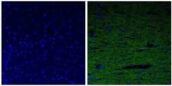
- Immunofluorescent analysis of the neurofilament light chain in paraffin-embedded rat brain tissue (right) compared to a negative control without primary antibody (left). Tissue sections were deparaffinized with xylene, and rehydrated with ethanol. To expose target proteins, antigen retrieval was performed using 10mM sodium citrate (pH 6.0) and microwaved for 8-15 min. Following antigen retrieval, tissues were washed with water and PBS, and then blocked in 0.3% BSA for 30 min at room temperature. Tissues were then probed with a neurofilament light chain monoclonal antibody (Product # 13-0400) in 0.3% BSA at a dilution of 1:20 for 1 hour at 37°C. Tissues were then incubated with a Goat anti-Mouse IgG (H+L) Secondary Antibody, DyLight 488 conjugate for 1 hour at 37°C (green). Nuclei (blue) were stained with DAPI. Images were taken at 40X magnification.

- Submitted by
- Invitrogen Antibodies (provider)
- Main image

- Experimental details
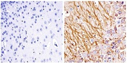
- Immunohistochemistry analysis of the neurofilament light chain showing staining in the filaments of paraffin-embedded human brain tissue (right) compared to a negative control without primary antibody (left). To expose target proteins, antigen retrieval was performed using 10mM sodium citrate (pH 6.0) and microwaved for 8-15 min. Following antigen retrieval, tissues were blocked in 3% H2O2-methanol for 15 min at room temperature, washed with ddH2O and PBS, and then probed with a Neurofilament light chain monoclonal antibody (Product # 13-0400) diluted in 3% BSA-PBS at a dilution of 1:20 overnight at 4°C in a humidified chamber. Tissues were washed extensively in PBST and detection was performed using an HRP-conjugated secondary antibody followed by colorimetric detection using a DAB kit. Tissues were counterstained with hematoxylin and dehydrated with ethanol and xylene to prep for mounting.

- Submitted by
- Invitrogen Antibodies (provider)
- Main image

- Experimental details
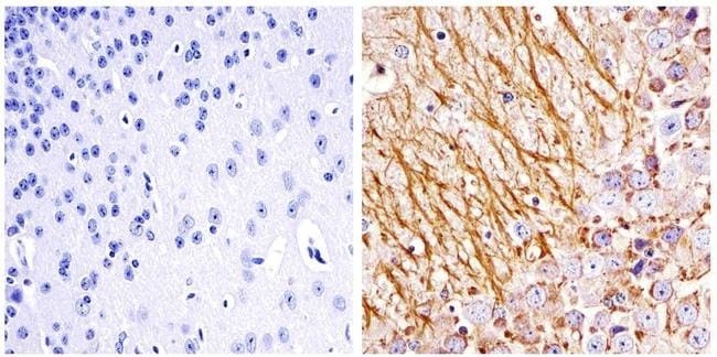
- Immunohistochemistry analysis of the neurofilament light chain showing staining in the filaments of paraffin-embedded rat brain tissue (right) compared to a negative control without primary antibody (left). To expose target proteins, antigen retrieval was performed using 10mM sodium citrate (pH 6.0) and microwaved for 8-15 min. Following antigen retrieval, tissues were blocked in 3% H2O2-methanol for 15 min at room temperature, washed with ddH2O and PBS, and then probed with a Neurofilament light chain monoclonal antibody (Product # 13-0400) diluted in 3% BSA-PBS at a dilution of 1:20 overnight at 4°C in a humidified chamber. Tissues were washed extensively in PBST and detection was performed using an HRP-conjugated secondary antibody followed by colorimetric detection using a DAB kit. Tissues were counterstained with hematoxylin and dehydrated with ethanol and xylene to prep for mounting.

- Submitted by
- Invitrogen Antibodies (provider)
- Main image

- Experimental details
- Immunofluorescent analysis of the neurofilament light chain in paraffin-embedded human brain tissue (right) compared to a negative control without primary antibody (left). Tissue sections were deparaffinized with xylene, and rehydrated with ethanol. To expose target proteins, antigen retrieval was performed using 10mM sodium citrate (pH 6.0) and microwaved for 8-15 min. Following antigen retrieval, tissues were washed with water and PBS, and then blocked in 0.3% BSA for 30 min at room temperature. Tissues were then probed with a neurofilament light chain monoclonal antibody (Product # 13-0400) in 0.3% BSA at a dilution of 1:20 for 1 hour at 37°C. Tissues were then incubated with a Goat anti-Mouse IgG (H+L) Secondary Antibody, DyLight 488 conjugate for 1 hour at 37°C (green). Nuclei (blue) were stained with DAPI. Images were taken at 40X magnification.

- Submitted by
- Invitrogen Antibodies (provider)
- Main image
- Experimental details
- Immunofluorescent analysis of the neurofilament light chain in paraffin-embedded rat brain tissue (right) compared to a negative control without primary antibody (left). Tissue sections were deparaffinized with xylene, and rehydrated with ethanol. To expose target proteins, antigen retrieval was performed using 10mM sodium citrate (pH 6.0) and microwaved for 8-15 min. Following antigen retrieval, tissues were washed with water and PBS, and then blocked in 0.3% BSA for 30 min at room temperature. Tissues were then probed with a neurofilament light chain monoclonal antibody (Product # 13-0400) in 0.3% BSA at a dilution of 1:20 for 1 hour at 37°C. Tissues were then incubated with a Goat anti-Mouse IgG (H+L) Secondary Antibody, DyLight 488 conjugate for 1 hour at 37°C (green). Nuclei (blue) were stained with DAPI. Images were taken at 40X magnification.

- Submitted by
- Invitrogen Antibodies (provider)
- Main image

- Experimental details
- Immunofluorescent analysis of the neurofilament light chain in paraffin-embedded mouse brain tissue (right) compared to a negative control without primary antibody (left). Tissue sections were deparaffinized with xylene, and rehydrated with ethanol. To expose target proteins, antigen retrieval was performed using 10mM sodium citrate (pH 6.0) and microwaved for 8-15 min. Following antigen retrieval, tissues were washed with water and PBS, and then blocked in 0.3% BSA for 30 min at room temperature. Tissues were then probed with a neurofilament light chain monoclonal antibody (Product # 13-0400) in 0.3% BSA at a dilution of 1:20 for 1 hour at 37°C. Tissues were then incubated with a Goat anti-Mouse IgG (H+L) Secondary Antibody, DyLight 488 conjugate for 1 hour at 37°C (green). Nuclei (blue) were stained with DAPI. Images were taken at 40X magnification.

- Submitted by
- Invitrogen Antibodies (provider)
- Main image
- Experimental details
- Immunohistochemistry analysis of the neurofilament light chain showing staining in the filaments of paraffin-embedded mouse brain tissue (right) compared to a negative control without primary antibody (left). To expose target proteins, antigen retrieval was performed using 10mM sodium citrate (pH 6.0) and microwaved for 8-15 min. Following antigen retrieval, tissues were blocked in 3% H2O2-methanol for 15 min at room temperature, washed with ddH2O and PBS, and then probed with a Neurofilament light chain monoclonal antibody (Product # 13-0400) diluted in 3% BSA-PBS at a dilution of 1:100 overnight at 4°C in a humidified chamber. Tissues were washed extensively in PBST and detection was performed using an HRP-conjugated secondary antibody followed by colorimetric detection using a DAB kit. Tissues were counterstained with hematoxylin and dehydrated with ethanol and xylene to prep for mounting.
Supportive validation
- Submitted by
- Invitrogen Antibodies (provider)
- Main image
- Experimental details
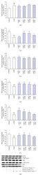
- Figure 6 Hippocampal GSK-3 beta , p-GSK-3 beta , CREB, p-CREB, BDNF, and NF-L protein levels in the corticosterone-induced mouse depression model were determined by Western blot analysis. The values of GSK-3 beta (a), CREB (c), BDNF (e), and NF-L (f) levels were normalized against the amount of beta -actin, while the values of p-GSK-3 beta (Ser9) (b) and p-CREB (Ser133) (d) were normalized against the amount of GSK-3 beta and CREB, respectively. ** P < 0.01 versus the CORT group, * P < 0.05 versus the CORT group, and ## P < 0.01 versus the control group.

- Submitted by
- Invitrogen Antibodies (provider)
- Main image

- Experimental details
- Figure 6 Hippocampal GSK-3 beta , p-GSK-3 beta , CREB, p-CREB, BDNF, and NF-L protein levels in the MS-induced mouse model were determined by Western blot analysis. The values of GSK-3 beta (a), CREB (c), BDNF (e), and NF-L (f) levels were normalized against the amount of beta -actin, while the values of p-GSK-3 beta (Ser9) (b) and p-CREB (Ser133) (d) were normalized against the amount of GSK-3 beta and CREB, respectively. * P < 0.01 versus the control group, # P < 0.01 versus the MS15 group.

- Submitted by
- Invitrogen Antibodies (provider)
- Main image
- Experimental details
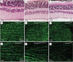
- Figure 2. Restoration of RGCs in Pou4f2 SpPou4f1/2/Z retinas. ( a-c ) H&E staining of retinas from mice at P60. ( d-f ) Immunofluorence staining of retinas from mice at P60 by anti-neurofilament heavy chain (SMI-32) antibody (green). ( g-i ) Immunofluorence staining of retinas from mice at P60 by anti-neurofilament ligh chain (NF-L) antibody (green). ( a , d , g ) Pou4f2 Z/+ control retinas. ( b , e,h ) Pou4f2 SpPou4f1/2/Z retinas. ( c , f , i ) Pou4f2 Z/Z retinas.

- Submitted by
- Invitrogen Antibodies (provider)
- Main image

- Experimental details
- Figure 1 Subcellular localization of Cep215 in P19 cells under differentiation. ( a ) Experimental scheme of glial differentiation of P19 cells. The cells were treated with retinoic acid (RA) for 4 days to induce embryoid body (EB) and cultured for up to 17 days. Neurogenesis occurs at the early stage of differentiation (D4-8) whereas gliogenesis occurs at the late stage of differentiation (D9-13). ( b ) P19 cells were treated with RA for differentiation and subjected to immunoblot analysis with antibodies specific to Nf68, Gfap and Gapdh. ( c ) The P19 cells at D17 were subjected to coimmunostaining analysis with antibodies specific to Cep215 (red) along with Tuj1 or Gfap (green). ( d-f ) Undifferentiated (UD) and differentiated (D12 and D15) P19 cells were subjected to immunoblot ( d ) and coimmunostaining ( e ) analyses with antibodies specific to Cep215, Gapdh and Gfap. ( f ) Intensities of the centrosomal Cep215 signals were measured. In case of D12 and D15, only Gfap-expressing cells were subjected to analysis. Greater than 90 cells per experimental group were estimated in three independent experiments. The statistical significance was analyzed using one-way ANOVA. * P < 0.05. ( c , e ) Nuclei were stained with DAPI (blue). Scale bars, 10 mum.

- Submitted by
- Invitrogen Antibodies (provider)
- Main image

- Experimental details
- Neurogenic potential in human brDFAT cells. ( A ) H&E staining of human BAT (interscapular) and WAT (subcutaneous) from human autopsy specimens (from subjects less than 3 months of age). ( B ) Expression of Uncoupling protein 1 (UCP1), Neurogenic Differentiation 1 (NeuroD1) and Neurofilament (NFL) in white and brown adipose tissue from human autopsy specimen as determined by qPCR (n = 3 cell preparations). P -values for statistically significant differences are indicated (unpaired t-tests). ( C ) Expression of CD105 and CD34 in human brDFAT cells as determined by FACS on day 7 of culture. ( D ) Expression of NFL (red) and NeuroD (green) in human brDFAT cells after 15 days of culture in neural induction medium, as visualized by immunofluorescence. DAPI (blue) was used to visualize nuclei. Representative images from 3-5 experiments.
