 Explore
 Validate
 Learn
Learn Western blot
 Immunocytochemistry
Antibody data
- Antibody Data
- Antigen structure
- References [0]
- Comments [0]
- Validations
- Western blot [1]
- ELISA [1]
- Chromatin Immunoprecipitation [1]
- Other assay [2]
Submit
Validation data
Reference
Comment
Report error
- Product number
- 49-1040 - Provider product page

- Provider
- Invitrogen Antibodies
- Product name
- Anti-Phospho-Tri-Methyl-Histone H3 (Ser10, Lys9) Polyclonal Antibody
- Antibody type
- Polyclonal
- Antigen
- Synthetic peptide
- Reactivity
- Human
- Host
- Rabbit
- Isotype
- IgG
- Vial size
- 100 µL
- Concentration
- Lot Dependent
- Storage
- -20° C, Avoid Freeze/Thaw Cycles
No comments: Submit comment
Supportive validation
- Submitted by
- Invitrogen Antibodies (provider)
- Main image

- Experimental details
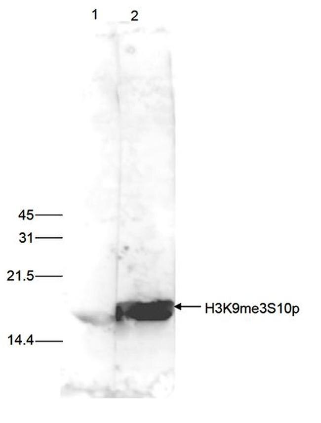
- HeLa cells were treated with colcemid to block the cell cycle in metaphase and 15 μg of histone extracts of these cells were analysed by Western blot with the anti-H3K9me3S10p antibody (Product # 49-1040) diluted 1:500 in TBS-Tween containing 5% skimmed milk. The position of the protein of interest is indicated on the right; the marker (in kDa) is shown on the left. The result of the Western analysis with the antibody is shown in lane 2; lane 1 shows the same analysis after incubation of the antibody with 5 nmol blocking peptide for 1 hour at room temperature.
Supportive validation
- Submitted by
- Invitrogen Antibodies (provider)
- Main image

- Experimental details
- To determine the titer, an ELISA was performed using a serial dilution of the anti-human H3K9me3S10p antibody (Product # 49-1040). The antigen used was a peptide containing the histone modification of interest. By plotting the absorbance against the antibody dilution, the titer of the antibody was estimated to be 1:87,000.

Supportive validation
- Submitted by
- Invitrogen Antibodies (provider)
- Main image

- Experimental details
- ChIP assays were performed using human HeLa cells treated with colcemid, the anti-H3K9me3S10p antibody (Product # 49-1040) and optimized PCR primer sets for qPCR. ChIP was performed using sheared chromatin from 10,000 cells per IP. A titration of the antibody consisting of 1, 5, and 10 μL per ChIP experiment was analysed. Additionally, the same titration was analysed after incubation of the antibody with 5 nmol blocking peptide for 1 hour at room temperature. IgG (5 μg/IP) was used as negative IP control. QPCR was performed with primers for the promoter of the active genes GAPDH and c-fos and for the heterochromatin marker Sat2. The figure shows the recovery, expressed as a % of input (the relative amount of immunoprecipitated DNA compared to input DNA after qPCR analysis).

Supportive validation
- Submitted by
- Invitrogen Antibodies (provider)
- Main image

- Experimental details
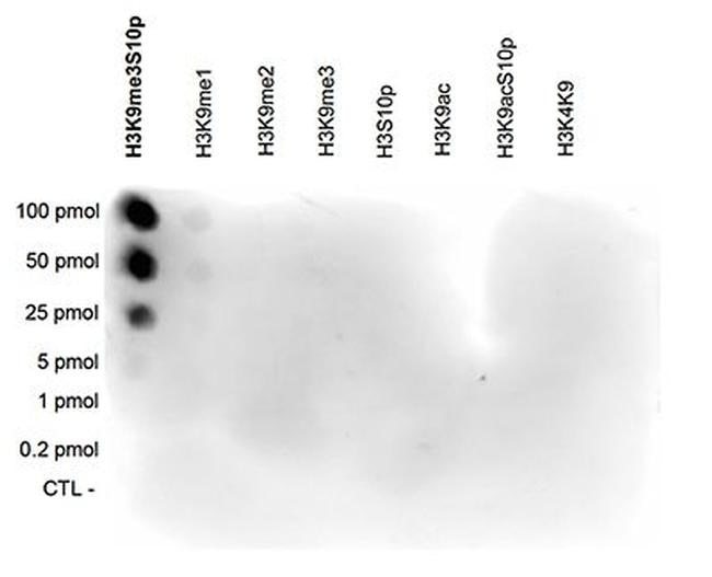
- A Dot Blot analysis was performed to test the cross reactivity of the anti-H3K9me3S10p antibody (Product # 49-1040) with peptides containing other modifications and unmodified sequences of histone H3. One hundred to 0.2 pmol of the peptide containing the respective histone modification were spotted on a membrane. The antibody was used at a dilution of 1:1,000. Figure 3 shows a high specificity of the antibody for the modification of interest.
- Submitted by
- Invitrogen Antibodies (provider)
- Main image

- Experimental details
- HeLa cells were stained with the anti-H3K9me3S10p antibody (Product # 49-1040) and with DAPI. Cells were fixed with 4% formaldehyde for 10’ and blocked with PBS/TX-100 containing 5% normal goat serum and 1% BSA. The cells were immunofluorescently labelled with the H3K9me3S10p antibody (left) diluted 1:500 in blocking solution followed by an anti-rabbit antibody conjugated to Alexa488. The middle panel shows staining of the nuclei with DAPI. A merge of the two stainings is shown on the right.
