 Explore
 Validate
 Learn
Learn Western blot
Antibody data
- Antibody Data
- Antigen structure
- References [0]
- Comments [0]
- Validations
- Western blot [1]
- ELISA [1]
- Immunocytochemistry [1]
- Immunoprecipitation [1]
- Other assay [2]
Submit
Validation data
Reference
Comment
Report error
- Product number
- TA347170 - Provider product page

- Provider
- OriGene
- Product name
- Rabbit Polyclonal H3K27me3S28p Antibody
- Antibody type
- Polyclonal
- Description
- Rabbit Polyclonal H3K27me3S28p Antibody
- Host
- Rabbit
- Conjugate
- Unconjugated
- Epitope
- HIST1H3A
- Isotype
- IgG
- Antibody clone number
- NULL
- Vial size
- 100 µl
- Concentration
- not determined
No comments: Submit comment
Supportive validation
- Submitted by
- OriGene (provider)
- Main image
- Experimental details
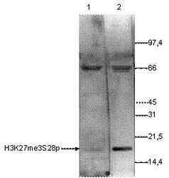
- WB using the antibody against H3K27 me3 S28 p diluted 1:250 in TBS-Tween containing 5% skimmed milk. The position of the protein of interest is indicated on the left; the marker (in kDa) is shown on the right. Lane 2 shows the result of the Western analysis with the antibody; lane 1 shows the same analysis after incubation of the antibody with 750 pmol blocking peptide for 1 hour at room temperature.
- Validation comment
- WB

Supportive validation
- Submitted by
- OriGene (provider)
- Main image
- Experimental details
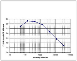
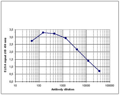
- Determination of the titer To determine the titer, an ELISA was performed using a serial dilution of the antibody against H3K27 me3 S28 p . The antigen used was a peptide containing the histone modifications of interest. By plotting the absorbance against the antibody dilution (Figure 2), the titer of the antibody was estimated to be 1:8,300.
- Validation comment
- ELISA
Supportive validation
- Submitted by
- OriGene (provider)
- Main image

- Experimental details
- HeLa asynchronous cells were fixed with formaldehyde, permeabilized with sodium citrate and Triton X100 and blocked with PBS with 2.5% BSA. A: cells were labelled with the antibody (1:200 and for 1 hr at RT) followed by goat anti-rabbit conjugated to DyLight 488. Figure 5B: the nuclei were stained with DAPI. Phosphorylation of H3 on serine 28 is increased during late G2 phase and reaches a maximum in metaphase cells. This may explain the different staining intensities of different cells.
- Validation comment
- IF

Supportive validation
- Submitted by
- OriGene (provider)
- Main image

- Experimental details
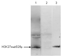
- HeLa cells were treated with colcemid to block the cell cycle in metaphase and were fixed with formaldehyde. Chromatin from 10,000 cells was sheared and used for immunoprecipitation (IP). IP was performed with 5ul of the ab. The IP'd proteins were analysed by WB with the antibody at 1:500 in TBS-Tween with 5% milk. Lane 1 shows the result of the IP; a negative IP control (no antibody added) and a positive control (sheared chromatin from 10,000 cells) are shown in lane 2 and 3.
- Validation comment
- IP
Supportive validation
- Submitted by
- OriGene (provider)
- Main image

- Experimental details
- ChIP assays using HeLa cells treated with colcemid: sheared chromatin from 10,000 cells. A titration of 1, 5 and 10 ul antibody per ChIP was performed after incubation with 5 nmol blocking peptide for 1 hr at RT. IgG (5 ug/IP) was used as negative control. qPCR primers were for the promoters of active genes c-fos and RPL30 and for inactive gene MYOD. Image shows the recovery, expressed as a % of input (the relative amount of IP'd DNA compared to input DNA after qPCR analysis).
- Validation comment
- Assay

- Submitted by
- OriGene (provider)
- Main image

- Experimental details
- A Dot Blot analysis was performed to test the cross reactivity of the antibody against H3K27 me3 S28 p with peptides containing other modifications of histone H3 and H4 and unmodified sequences from histone H3. One hundred to 0.2 pmol of the peptides were spotted on a membrane. The antibody was used at a dilution of 1:20,000. Image shows a high specificity of the antibody for the peptide containing the modifications of interest.
- Validation comment
- DB
