 Explore
 Validate
 Learn
Learn Western blot
 Immunoprecipitation
Antibody data
- Antibody Data
- Antigen structure
- References [1]
- Comments [0]
- Validations
- Western blot [6]
- Flow cytometry [3]
Submit
Validation data
Reference
Comment
Report error
- Product number
- MA1-19197 - Provider product page

- Provider
- Invitrogen Antibodies
- Product name
- LCK Monoclonal Antibody (LCK-01)
- Antibody type
- Monoclonal
- Antigen
- Synthetic peptide
- Description
- This antibody recognizes defined epitope (aa 22-36) of Lck, a 56 kDa Src-family protein tyrosine kinase (intracellular antigen). This antibody will not cross-react with mouse.
- Reactivity
- Human
- Host
- Mouse
- Isotype
- IgG
- Antibody clone number
- LCK-01
- Vial size
- 100 µg
- Concentration
- 1 mg/mL
- Storage
- 4° C, do not freeze
Submitted references The phosphoproteome of human Jurkat T cell clones upon costimulation with anti-CD3/anti-CD28 antibodies.
Nguyen TD, Carrascal M, Vidal-Cortes O, Gallardo O, Casas V, Gay M, Phan VC, Abian J
Journal of proteomics 2016 Jan 10;131:190-198
Journal of proteomics 2016 Jan 10;131:190-198
No comments: Submit comment
Supportive validation
- Submitted by
- Invitrogen Antibodies (provider)
- Main image

- Experimental details
- Western blot analysis of LCK using a monoclonal antibody (Product # MA1-19197).

- Submitted by
- Invitrogen Antibodies (provider)
- Main image

- Experimental details
- Western blot analysis of LCK using a monoclonal antibody (Product # MA1-19197).

- Submitted by
- Invitrogen Antibodies (provider)
- Main image

- Experimental details
- Western blotting analysis (reducing conditions) of human Lck in whole cell lysate using anti-human Lck (LCK-01) Monoclonal antibody (Product # MA1-19197). Lane 1: J. CaM-1.6 cell line (a mutant derivate of the JURKAT cell line) transfected with Lck; Lane 2: HEK-293T cell line transfected with Lck; Lane 3: HEK-293T cell line (non-transfected).

- Submitted by
- Invitrogen Antibodies (provider)
- Main image

- Experimental details
- Knockdown of LCK was achieved by transfecting Jurkat with LCK specific siRNAs (Silencer® select Product # s8108, s8107). Western blot analysis (Fig. a) was performed using whole cell extracts from the LCK knockdown cells (Lane 3), non-specific scrambled siRNA transfected cells (Lane 2) and untransfected cells (Lane 1). The blot was probed with LCK Monoclonal Antibody (LCK-01) (Product # MA1-19197, 1:1000 dilution) and Goat anti-Mouse IgG (H+L) Superclonal™ Recombinant Secondary Antibody, HRP (Product # A28177, 1:4000 dilution). Densitometric analysis of this western blot is shown in histogram (Fig. b). Decrease in signal upon siRNA mediated knock down confirms that antibody is specific to LCK.

- Submitted by
- Invitrogen Antibodies (provider)
- Main image

- Experimental details
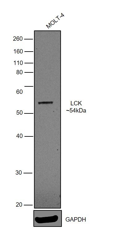
- Western blot was performed using Anti-LCK Monoclonal Antibody (LCK-01) (Product # MA1-19197) and a 54kDa band corresponding to LCK was observed the tested cell model. Whole cell lysates (30ug lysate) of MOLT-4 (Lane 1) were electrophoresed using Novex® NuPAGE® 4-12 % Bis-Tris gel (Product # NP0322BOX). Resolved proteins were then transferred onto a nitrocellulose membrane (Product # IB23001) by iBlot® 2 Dry Blotting System (Product # IB21001). The bot was probed with the primary antibody (1:1000 dilution) detected by chemiluminescence with Goat anti-Mouse IgG (H+L) Superclonal™ Recombinant Secondary Antibody, HRP (Product # A28177, 1:4000 dilution) using the iBright FL 1000 (Product # A32752). Chemiluminescent detection was performed using Novex® ECL Chemiluminescent Substrate Reagent Kit (Product # WP20005).
Supportive validation
- Submitted by
- Invitrogen Antibodies (provider)
- Main image

- Experimental details
- Flow cytometry intracellular staining pattern of human peripheral whole blood using anti-LCK (LCK-01) purified Monoclonal antibody (Product # MA1-19197) (concentration in sample 9 µg/mL, GAM APC).

- Submitted by
- Invitrogen Antibodies (provider)
- Main image

- Experimental details
- Separation of human CD3 positive LCK positive human lymphocytes (red-filled) from neutrophil granulocytes (black-dashed) in flow cytometry analysis (intracellular staining) of peripheral whole blood stained using anti-LCK (LCK-01) purified Monoclonal antibody (Product # MA1-19197) (concentration in sample 9 µg/mL, GAM APC).

- Submitted by
- Invitrogen Antibodies (provider)
- Main image

- Experimental details
- Flow cytometry multicolor intracellular staining of human peripheral whole blood stained using anti-LCK (LCK-01) purified Monoclonal antibody (Product # MA1-19197) (concentration in sample 9 µg/mL, GAM APC) and anti-human CD3 (UCHT1) Pacific Blue™ using a dilution of 20 µL reagent/100 µL of peripheral whole blood.
