 Explore
 Validate
 Learn
Learn Western blot
Antibody data
- Antibody Data
- Antigen structure
- References [5]
- Comments [0]
- Validations
- Western blot [1]
- Immunocytochemistry [1]
Submit
Validation data
Reference
Comment
Report error
- Product number
- MAB7500 - Provider product page

- Provider
- R&D Systems
- Product name
- Human Phospho-Lck (Y394) Antibody
- Antibody type
- Monoclonal
- Description
- Protein A or G purified from hybridoma culture supernatant. Detects human Lck when phosphorylated at Y394. Due to high sequence homology, this antibody may cross-react with phosphorylated members of the Src family.
- Reactivity
- Human
- Host
- Mouse
- Conjugate
- Unconjugated
- Antigen sequence
P06239- Isotype
- IgG
- Antibody clone number
- 755103
- Vial size
- 100 ug
- Concentration
- LYOPH
- Storage
- Use a manual defrost freezer and avoid repeated freeze-thaw cycles. 12 months from date of receipt, -20 to -70 °C as supplied. 1 month, 2 to 8 °C under sterile conditions after reconstitution. 6 months, -20 to -70 °C under sterile conditions after reconstitution.
Submitted references Nuclear pore complex-mediated modulation of TCR signaling is required for naïve CD4+ T cell homeostasis.
Characterization of the effects of immunomodulatory drug fingolimod (FTY720) on human T cell receptor signaling pathways.
TAOK3 Regulates Canonical TCR Signaling by Preventing Early SHP-1-Mediated Inactivation of LCK.
Hepatitis C virus infection inhibits a Src-kinase regulatory phosphatase and reduces T cell activation in vivo.
T cell Ig and mucin domain-containing protein 3 is recruited to the immune synapse, disrupts stable synapse formation, and associates with receptor phosphatases.
Borlido J, Sakuma S, Raices M, Carrette F, Tinoco R, Bradley LM, D'Angelo MA
Nature immunology 2018 Jun;19(6):594-605
Nature immunology 2018 Jun;19(6):594-605
Characterization of the effects of immunomodulatory drug fingolimod (FTY720) on human T cell receptor signaling pathways.
Baer A, Colon-Moran W, Bhattarai N
Scientific reports 2018 Jul 19;8(1):10910
Scientific reports 2018 Jul 19;8(1):10910
TAOK3 Regulates Canonical TCR Signaling by Preventing Early SHP-1-Mediated Inactivation of LCK.
Ormonde JVS, Li Z, Stegen C, Madrenas J
Journal of immunology (Baltimore, Md. : 1950) 2018 Dec 1;201(11):3431-3442
Journal of immunology (Baltimore, Md. : 1950) 2018 Dec 1;201(11):3431-3442
Hepatitis C virus infection inhibits a Src-kinase regulatory phosphatase and reduces T cell activation in vivo.
Bhattarai N, McLinden JH, Xiang J, Mathahs MM, Schmidt WN, Kaufman TM, Stapleton JT
PLoS pathogens 2017 Feb;13(2):e1006232
PLoS pathogens 2017 Feb;13(2):e1006232
T cell Ig and mucin domain-containing protein 3 is recruited to the immune synapse, disrupts stable synapse formation, and associates with receptor phosphatases.
Clayton KL, Haaland MS, Douglas-Vail MB, Mujib S, Chew GM, Ndhlovu LC, Ostrowski MA
Journal of immunology (Baltimore, Md. : 1950) 2014 Jan 15;192(2):782-91
Journal of immunology (Baltimore, Md. : 1950) 2014 Jan 15;192(2):782-91
No comments: Submit comment
Supportive validation
- Submitted by
- R&D Systems (provider)
- Main image
- Experimental details
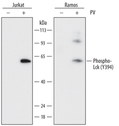
- Detection of Human Phospho-Lck (Y394) by Western Blot. Western blot shows lysates of Jurkat human acute T cell leukemia cell line and Ramos human Burkitt's lymphoma cell line untreated (-) or treated (+) with 1 mM Pervanadate (PV) for 5 minutes. PVDF membrane was probed with 0.1 µg/mL of Mouse Anti-Human Phospho-Lck (Y394) Monoclonal Antibody (Catalog # MAB7500) followed by HRP-conjugated Anti-Mouse IgG Secondary Antibody (Catalog # HAF018). A specific band was detected for Phospho-Lck (Y394) at approximately 56 kDa (as indicated). This experiment was conducted under reducing conditions and using Immunoblot Buffer Group 1.

Supportive validation
- Submitted by
- R&D Systems (provider)
- Main image

- Experimental details
- Phospho-Lck (Y394) in Jurkat Human Cell Line. Lck phosphorylated at Y394 was detected in immersion fixed Jurkat human acute T cell leukemia cells treated with (upper panel) or without (lower panel) Pervanadate using Mouse Anti-Human Phospho-Lck (Y394) Monoclonal Antibody (Catalog # MAB7500) at 10 µg/mL for 3 hours at room temperature. Cells were stained using the NorthernLights™ 557-conjugated Anti-Mouse IgG Secondary Antibody (red; Catalog # NL007) and counterstained with DAPI (blue). Specific staining was localized to cell surfaces and cytoplasm. View our protocol for Fluorescent ICC Staining of Non-adherent Cells.
