 Explore
 Validate
 Learn
Learn Western blot
Antibody data
- Antibody Data
- Antigen structure
- References [3]
- Comments [0]
- Validations
- Western blot [1]
- Immunocytochemistry [1]
- Immunohistochemistry [1]
- Flow cytometry [1]
Submit
Validation data
Reference
Comment
Report error
- Product number
- GTX16613 - Provider product page

- Provider
- GeneTex
- Product name
- Orai1 antibody
- Antibody type
- Polyclonal
- Reactivity
- Human, Mouse, Rat
- Host
- Rabbit
Submitted references Termination of T cell priming relies on a phase of unresponsiveness promoting disengagement from APCs and T cell division.
Store-operated Ca2+ Entry Facilitates the Lipopolysaccharide-induced Cyclooxygenase-2 Expression in Gastric Cancer Cells.
STIM2 enhances receptor-stimulated Ca²⁺ signaling by promoting recruitment of STIM1 to the endoplasmic reticulum-plasma membrane junctions.
Bohineust A, Garcia Z, Beuneu H, Lemaître F, Bousso P
The Journal of experimental medicine 2018 May 7;215(5):1481-1492
The Journal of experimental medicine 2018 May 7;215(5):1481-1492
Store-operated Ca2+ Entry Facilitates the Lipopolysaccharide-induced Cyclooxygenase-2 Expression in Gastric Cancer Cells.
Wong JH, Ho KH, Nam S, Hsu WL, Lin CH, Chang CM, Wang JY, Chang WC
Scientific reports 2017 Oct 16;7(1):12813
Scientific reports 2017 Oct 16;7(1):12813
STIM2 enhances receptor-stimulated Ca²⁺ signaling by promoting recruitment of STIM1 to the endoplasmic reticulum-plasma membrane junctions.
Ong HL, de Souza LB, Zheng C, Cheng KT, Liu X, Goldsmith CM, Feske S, Ambudkar IS
Science signaling 2015 Jan 13;8(359):ra3
Science signaling 2015 Jan 13;8(359):ra3
No comments: Submit comment
Supportive validation
- Submitted by
- GeneTex (provider)
- Main image

- Experimental details
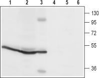
- Anti-Orai1_(extracellular) - Western blot analysis of rat spleen (lanes 1 and4), rat pancreas (lanes 2 and 5) and mouse B-cell lymphoma (WEHI) (lanes 3 and 6) lysates: 1-3. Anti-Orai1 (extracellular) antibody, (1:200). 4-6. Anti-Orai1 (extracellular) antibody, preincubated with the control peptide antigen.
Supportive validation
- Submitted by
- GeneTex (provider)
- Main image

- Experimental details
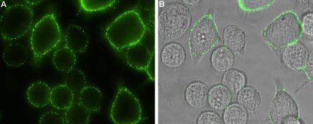
- Anti-Orai1_(extracellular) - Expression of Orai1 in rat RBL cells Immunocytochemical staining of intact living rat basophilic leukemia (RBL) cells. A. Extracellular staining of cells using Anti-Orai1 (extracellular) antibody, (1:100) followed by goat anti-rabbit-AlexaFluor-488 secondary antibody (green). B. Merge of A and Live view of the cells.
Supportive validation
- Submitted by
- GeneTex (provider)
- Main image

- Experimental details
- Anti-Orai1_(extracellular) - Expression of Orai1 in rat pancreas Immunohistochemical staining of rat pancreas paraffin embedded sections using Anti-Orai1 (extracellular) antibody, (1:50). Orai1 (brown) is expressed in acinar cells. Hematoxilin is used as the counterstain.

Supportive validation
- Submitted by
- GeneTex (provider)
- Main image

- Experimental details
- Anti-Orai1_(extracellular) - Indirect flow cytometry analysis of WEHI living cells: Black:Unstained cells. Green:Cells + Anti-Orai1 (extracellular) antibody, (10 £gg/0.5 x 106 cells).
