Explore
 Validate
 Learn
Learn Western blot
Antibody data
- Antibody Data
- Antigen structure
- References [0]
- Comments [0]
- Validations
- Western blot [1]
- Immunocytochemistry [1]
- Immunohistochemistry [1]
Submit
Validation data
Reference
Comment
Report error
- Product number
- TA328818 - Provider product page

- Provider
- OriGene
- Product name
- Rabbit Polyclonal Anti-GABA(A) Theta Receptor (extracellular)
- Antibody type
- Polyclonal
- Description
- Rabbit Polyclonal Anti-GABA(A) Theta Receptor (extracellular)
- Host
- Rabbit
- Conjugate
- Unconjugated
- Epitope
- Gabrq
- Antibody clone number
- NULL
- Vial size
- 200 µl
- Concentration
- NULL
No comments: Submit comment
Supportive validation
- Submitted by
- OriGene (provider)
- Main image

- Experimental details
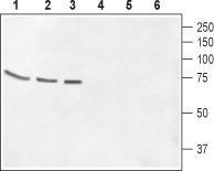
- Western blot analysis of rat brain membrane (lanes 1 and 4), mouse brain membrane (lanes 2 and 5) and CCF-STGI cell lysate (lanes 3 and 6): 1-3. Anti-GABA(A) ? Receptor (extracellular) antibody, (1:500). 4-6. Anti-GABA(A) ? Receptor (extracellular) antibody, preincubated with the control peptide antigen.
- Validation comment
- WB
Supportive validation
- Submitted by
- OriGene (provider)
- Main image

- Experimental details
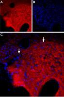
- Expression of GABA(A) ? receptor in rat PC12 cells. Immunocytochemical staining of intact living rat Pheochromocytoma (PC12) cells. A. Extracellular staining of cells using Anti-GABA(A) ? Receptor (extracellular) antibody , (1:50), (red). B. Merge of A with the live view of the cell.
- Validation comment
- IF

Supportive validation
- Submitted by
- OriGene (provider)
- Main image

- Experimental details
- Expression of of GABA(A) ? Receptor in rat hypothalamus. Immunohistochemical staining of rat hypothalamus using Anti-GABA(A) ? Receptor (extracellular) antibody . A. GABA(A) ? Receptor staining (red) is detected in the mammillary nucleus which is part of the posterior hypothalamus (arrows show the border of the nucleus). B. Nuclear staining using DAPI as the counterstain (blue). C. Merge images of A and B.
- Validation comment
- IHC