Explore
 Validate
 Learn
Learn Western blot
 Immunohistochemistry
Antibody data
- Antibody Data
- Antigen structure
- References [0]
- Comments [0]
- Validations
- Western blot [1]
Submit
Validation data
Reference
Comment
Report error
- Product number
- A05636-1 - Provider product page

- Provider
- Boster Biological Technology
- Product name
- Anti-NARG1/NAA15 Antibody Picoband™
- Antibody type
- Polyclonal
- Description
- Polyclonal antibody for NARG1/NAA15 detection. Host: Rabbit.Size: 100μg/vial. Tested applications: IHC-P. Reactive species: Human. NARG1/NAA15 information: Molecular Weight: 101272 MW; Subcellular Localization: Cytoplasm. Nucleus. Mainly cytoplasmic, nuclear in some cases. Present in the free cytosolic and cytoskeleton-bound polysomes, but not in the membrane-bound polysomes; Tissue Specificity: Expressed at high levels in testis and in ocular endothelial cells. Also found in brain (corpus callosum), heart, colon, bone marrow and at lower levels in most adult tissues, including thyroid, liver, pancreas, mammary and salivary glands, lung, ovary, urogenital system and upper gastrointestinal tract. Overexpressed in gastric cancer, in papillary thyroid carcinomas and in a Burkitt lymphoma cell line (Daudi). Specifically suppressed in abnormal proliferating blood vessels in eyes of patients with proliferative diabetic retinopathy.
- Reactivity
- Human, Mouse
- Host
- Rabbit
- Vial size
- 100μg/vial
- Concentration
- Add 0.2ml of distilled water will yield a concentration of 500ug/ml.
- Storage
- At -20°C for one year. After reconstitution, at 4°C for one month. It can also be aliquoted and stored frozen at -20°C for a longer time. Avoid repeated freezing and thawing.
- Handling
- Add 0.2ml of distilled water will yield a concentration of 500ug/ml.
No comments: Submit comment
Supportive validation
- Submitted by
- Boster Biological Technology (provider)
- Main image
- Experimental details
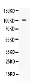
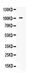
- Western blot analysis of NARG1 expression in 293T whole cell lysates (lane 1). NARG1 at 101KD was detected using rabbit anti- NARG1 Antigen Affinity purified polyclonal antibody (Catalog #A05636-1) at 0.5 μg/mL. The blot was developed using chemiluminescence (ECL) method (Catalog # EK1002).
- Additional image