Explore
 Validate
 Learn
Learn701796-20UG
antibody from Invitrogen Antibodies
Targeting: H4C14
H4/n, H4F2, H4FN, HIST2H4, HIST2H4A
 Western blot
Antibody data
- Antibody Data
- Antigen structure
- References [1]
- Comments [0]
- Validations
- Western blot [2]
- Immunocytochemistry [1]
- Other assay [3]
Submit
Validation data
Reference
Comment
Report error
- Product number
- 701796-20UG - Provider product page

- Provider
- Invitrogen Antibodies
- Product name
- H4K8ac Recombinant Rabbit Monoclonal Antibody (8H5L4), ChIP-Verified
- Antibody type
- Monoclonal
- Antigen
- Synthetic peptide
- Description
- Since it is highly conserved across species, the antibody may react with many other species.
- Antibody clone number
- 8H5L4
- Concentration
- 0.5 mg/mL
Submitted references Interferon regulatory factor 1 and a variant of heterogeneous nuclear ribonucleoprotein L coordinately silence the gene for adhesion protein CEACAM1.
Dery KJ, Silver C, Yang L, Shively JE
The Journal of biological chemistry 2018 Jun 15;293(24):9277-9291
The Journal of biological chemistry 2018 Jun 15;293(24):9277-9291
No comments: Submit comment
Supportive validation
- Submitted by
- Invitrogen Antibodies (provider)
- Main image

- Experimental details
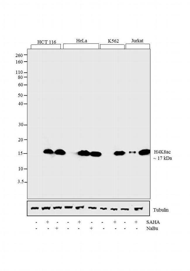
- Western blot analysis was performed on whole cell extracts (30 µg lysate) of HCT 116 (Lane1), HCT 116 treated with SAHA (0.5 uM/ 24 hours) (Lane 2), HCT 116 treated with sodium butyrate (5mM/ 24hours) (Lane 3), HeLa (Lane 4), HeLa treated with SAHA (0.5 uM/ 24 hours) (Lane 5), HeLa treated with sodium butyrate (5mM/ 24 hours) (Lane 6), K562 (Lane 7), K562 treated with SAHA (0.5 uM/ 24 hours) (Lane 8), Jurkat (Lane 9) and Jurkat treated with SAHA (0.5 uM/ 24 hours) (Lane 10). The blots were probed with Anti-Histone H4K8ac Recombinant Rabbit Monoclonal Antibody (Product # 701796 0.5-1 µg/mL) and detected by chemiluminescence using Goat anti-Rabbit IgG (H+L) Superclonal Secondary Antibody, HRP conjugate (Product # A27036, 0.4 µg/mL, 1:2500 dilution). A clear 17kDa band corresponding to Histone H4K8ac was observed across cell lines tested. Known quantity of protein samples were electrophoresed using Novex®NuPAGE®4-12% Bis-Tris gel (Product # NP0321BOX), XCell SureLock Electrophoresis System (Product # EI0002), and Novex® Sharp Pre-Stained Protein Standard (Product # LC5800). Resolved proteins were then transferred onto a nitrocellulose membrane with iBlot® Dry Blotting System (Product # IB21001). The membrane was probed with the relevant primary and secondary antibody following blocking with 5% skimmed milk. Chemiluminescent detection was performed using Pierce™ ECL Western blotting Substrate (Product # 32106).
- Submitted by
- Invitrogen Antibodies (provider)
- Main image
- Experimental details
- Western blot analysis was performed on whole cell extracts (30 µg lysate) of HCT 116 (Lane1), HCT 116 treated with SAHA (0.5 uM/ 24 hours) (Lane 2), HCT 116 treated with sodium butyrate (5mM/ 24hours) (Lane 3), HeLa (Lane 4), HeLa treated with SAHA (0.5 uM/ 24 hours) (Lane 5), HeLa treated with sodium butyrate (5mM/ 24 hours) (Lane 6), K562 (Lane 7), K562 treated with SAHA (0.5 uM/ 24 hours) (Lane 8), Jurkat (Lane 9) and Jurkat treated with SAHA (0.5 uM/ 24 hours) (Lane 10). The blots were probed with Anti-Histone H4K8ac Recombinant Rabbit Monoclonal Antibody (Product # 701796 0.5-1 µg/mL) and detected by chemiluminescence using Goat anti-Rabbit IgG (H+L) Superclonal Secondary Antibody, HRP conjugate (Product # A27036, 0.4 µg/mL, 1:2500 dilution). A clear 17kDa band corresponding to Histone H4K8ac was observed across cell lines tested. Known quantity of protein samples were electrophoresed using Novex®NuPAGE®4-12% Bis-Tris gel (Product # NP0321BOX), XCell SureLock Electrophoresis System (Product # EI0002), and Novex® Sharp Pre-Stained Protein Standard (Product # LC5800). Resolved proteins were then transferred onto a nitrocellulose membrane with iBlot® Dry Blotting System (Product # IB21001). The membrane was probed with the relevant primary and secondary antibody following blocking with 5% skimmed milk. Chemiluminescent detection was performed using Pierce™ ECL Western blotting Substrate (Product # 32106).

Supportive validation
- Submitted by
- Invitrogen Antibodies (provider)
- Main image

- Experimental details
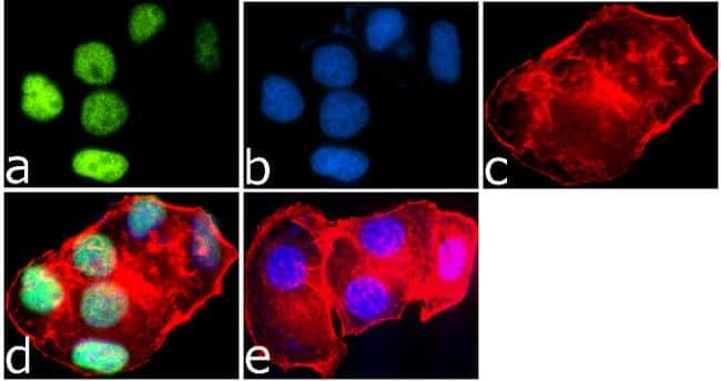
- Immunofluorescence was performed on MCF7 cells treated with SAHA (0.5 uM/18 hours). These cells were permeabilized and fixed for detection of Histone H4K8ac using Anti-Histone H4K8ac Recombinant Rabbit Monoclonal Antibody (Product # 701796, 1 µg/mL) and labeled with Goat anti-Rabbit IgG (H+L) Superclonal Secondary Antibody, Alexa Fluor® 488 conjugate (Product # A27034, 0.4 µg/mL, 1:2500). Panel a) shows representative cells that were stained for detection and localization of Histone H4K8ac protein (green), Panel b) is stained for Nuclei (blue) using SlowFade® Gold Antifade Mountant with DAPI (Product # S36938, 1:50). Panel c) represents cytoskeletal F-actin staining using Alexa Fluor® 594 Phalloidin (Product # A12381, 1:200). Panel d) is a composite image of Panels a, b and c clearly demonstrating nuclear localization of Histone H4K8ac. Panel e) represents control cells with no primary antibody to assess background.
Supportive validation
- Submitted by
- Invitrogen Antibodies (provider)
- Main image

- Experimental details
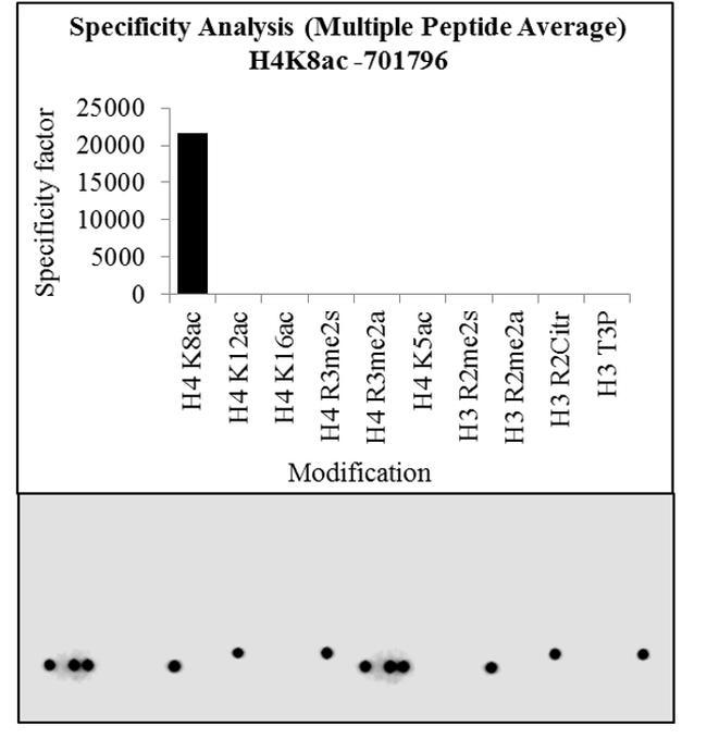
- Antibody specificity for modified targets can be established using peptide arrays by quantifying detection of the target protein along with closely related proteins. Peptide array of Histone H4K8Ac using Anti-Acetyl-Histone H4 (Lys8) Recombinant Rabbit Monoclonal Antibody: An array of the specific peptide and other relevant peptides when tested using Anti-Acetyl-Histone H4 (Lys8) Recombinant Rabbit Monoclonal Antibody (Product # 701796), showed that the Histone H4K8Ac modification was specifically recognized by the antibody.
- Submitted by
- Invitrogen Antibodies (provider)
- Main image
- Experimental details
- Antibody specificity for modified targets can be established using peptide arrays by quantifying detection of the target protein along with closely related proteins. Peptide array of Histone H4K8Ac using Anti-Acetyl-Histone H4 (Lys8) Recombinant Rabbit Monoclonal Antibody: An array of the specific peptide and other relevant peptides when tested using Anti-Acetyl-Histone H4 (Lys8) Recombinant Rabbit Monoclonal Antibody (Product # 701796), showed that the Histone H4K8Ac modification was specifically recognized by the antibody.

- Submitted by
- Invitrogen Antibodies (provider)
- Main image

- Experimental details
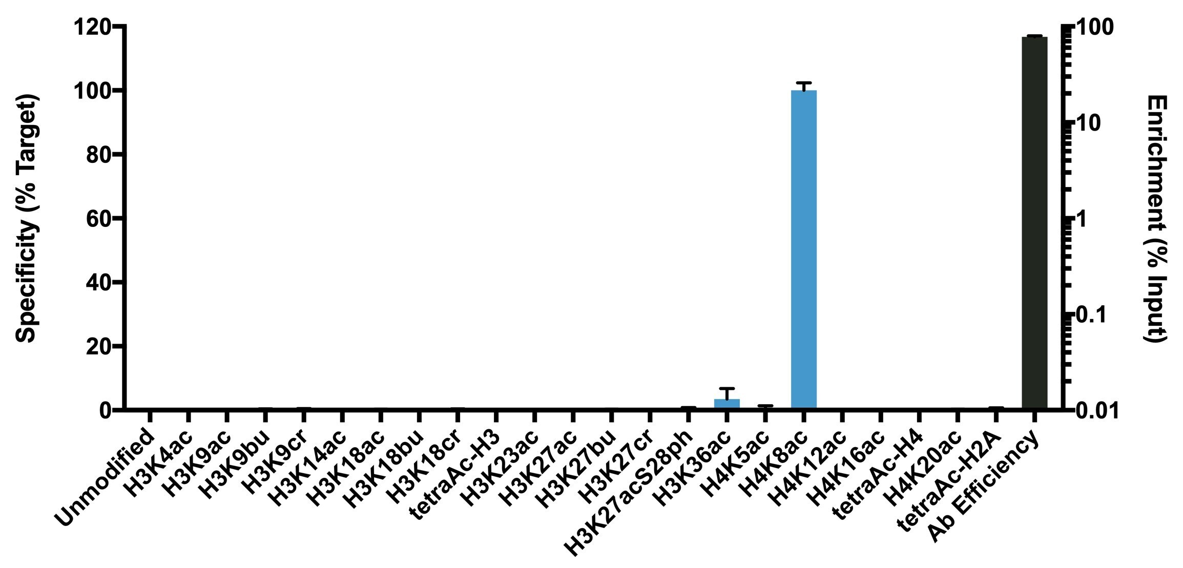
- Antibody specificity was demonstrated by detection of enrichment of the targeted histone modification using SNAP-ChIP™ Spike-in, a proprietary technology developed by EpiCypher™. SNAP-ChIP™ spike-in was performed using H4K8ac Recombinant Rabbit Monoclonal Antibody (Product # 701796) and H4K8ac was enriched compared to the other histone modifications in the SNAP-ChIP™ K-AcylStat Panel (Product # A47358).