Explore
 Validate
 Learn
Learn Western blot
 Immunocytochemistry
Antibody data
- Antibody Data
- Antigen structure
- References [5]
- Comments [0]
- Validations
- Immunocytochemistry [1]
- Immunohistochemistry [2]
- Flow cytometry [1]
Submit
Validation data
Reference
Comment
Report error
- Product number
- MAB1129-100 - Provider product page

- Provider
- R&D Systems
- Product name
- Human ErbB2/Her2 Antibody
- Antibody type
- Monoclonal
- Description
- Protein A or G purified from hybridoma culture supernatant.
- Reactivity
- Human
- Host
- Mouse
- Conjugate
- Unconjugated
- Antigen sequence
P04626- Isotype
- IgG
- Antibody clone number
- 191924
- Vial size
- 100 ug
- Concentration
- LYOPH
- Storage
- Use a manual defrost freezer and avoid repeated freeze-thaw cycles. 12 months from date of receipt, -20 to -70 °C as supplied. 1 month, 2 to 8 °C under sterile conditions after reconstitution. 6 months, -20 to -70 °C under sterile conditions after reconstitution.
Submitted references Antibody-drug conjugate T-DM1 treatment for HER2+ breast cancer induces ROR1 and confers resistance through activation of Hippo transcriptional coactivator YAP1.
Detection of Circulating Tumour Cells in Urothelial Cancers and Clinical Correlations: Comparison of Two Methods.
CRISPR-Cas9 epigenome editing enables high-throughput screening for functional regulatory elements in the human genome.
HER2 and EGFR Overexpression Support Metastatic Progression of Prostate Cancer to Bone.
Microbead arrays for the analysis of ErbB receptor tyrosine kinase activation and dimerization in breast cancer cells.
Islam SS, Uddin M, Noman ASM, Akter H, Dity NJ, Basiruzzman M, Uddin F, Ahsan J, Annoor S, Alaiya AA, Al-Alwan M, Yeger H, Farhat WA
EBioMedicine 2019 May;43:211-224
EBioMedicine 2019 May;43:211-224
Detection of Circulating Tumour Cells in Urothelial Cancers and Clinical Correlations: Comparison of Two Methods.
Fina E, Necchi A, Bottelli S, Reduzzi C, Pizzamiglio S, Iacona C, Daidone MG, Verderio P, Cappelletti V
Disease markers 2017;2017:3414910
Disease markers 2017;2017:3414910
CRISPR-Cas9 epigenome editing enables high-throughput screening for functional regulatory elements in the human genome.
Klann TS, Black JB, Chellappan M, Safi A, Song L, Hilton IB, Crawford GE, Reddy TE, Gersbach CA
Nature biotechnology 2017 Jun;35(6):561-568
Nature biotechnology 2017 Jun;35(6):561-568
HER2 and EGFR Overexpression Support Metastatic Progression of Prostate Cancer to Bone.
Day KC, Lorenzatti Hiles G, Kozminsky M, Dawsey SJ, Paul A, Broses LJ, Shah R, Kunja LP, Hall C, Palanisamy N, Daignault-Newton S, El-Sawy L, Wilson SJ, Chou A, Ignatoski KW, Keller E, Thomas D, Nagrath S, Morgan T, Day ML
Cancer research 2017 Jan 1;77(1):74-85
Cancer research 2017 Jan 1;77(1):74-85
Microbead arrays for the analysis of ErbB receptor tyrosine kinase activation and dimerization in breast cancer cells.
Khan IH, Zhao J, Ghosh P, Ziman M, Sweeney C, Kung HJ, Luciw PA
Assay and drug development technologies 2010 Feb;8(1):27-36
Assay and drug development technologies 2010 Feb;8(1):27-36
No comments: Submit comment
Supportive validation
- Submitted by
- R&D Systems (provider)
- Main image
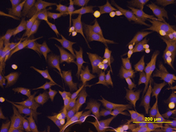
- Experimental details
- ErbB2/Her2 in MDA-MB-231 Human Cell Line. ErbB2/Her2 was detected in immersion fixed MDA-MB-231 human breast cancer cell line using Mouse Anti-Human ErbB2/Her2 Monoclonal Antibody (Catalog # MAB1129) at 10 µg/mL for 3 hours at room temperature. Cells were stained using the NorthernLights™ 557-conjugated Anti-Mouse IgG Secondary Antibody (yellow; Catalog # NL007) and counterstained with DAPI (blue). View our protocol for Fluorescent ICC Staining of Cells on Coverslips.
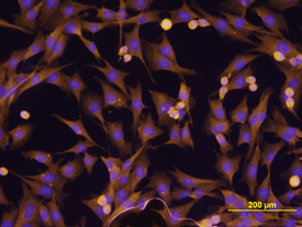
Supportive validation
- Submitted by
- R&D Systems (provider)
- Main image
- Experimental details
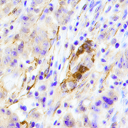
- ErbB2/Her2 in Human Stomach. ErbB2/Her2 was detected in immersion fixed paraffin-embedded sections of human stomach using Mouse Anti-Human ErbB2/Her2 Monoclonal Antibody (Catalog # MAB1129) at 15 µg/mL overnight at 4 °C. Tissue was stained using the Anti-Mouse HRP-DAB Cell & Tissue Staining Kit (brown; Catalog # CTS002) and counterstained with hematoxylin (blue). Specific staining was localized to plasma membrane/cytoplasm in glans and villi. View our protocol for Chromogenic IHC Staining of Paraffin-embedded Tissue Sections.

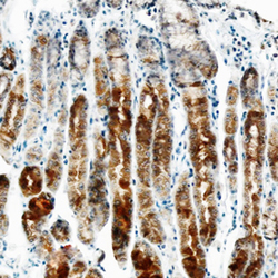
- Submitted by
- R&D Systems (provider)
- Main image
- Experimental details
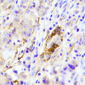
- ErbB2/Her2 in Human Breast Cancer Tissue. ErbB2/Her2 was detected in immersion fixed paraffin-embedded sections of human breast cancer tissue using Mouse Anti-Human ErbB2/Her2 Monoclonal Antibody (Catalog # MAB1129) at 15 µg/mL overnight at 4 °C. Tissue was stained using the Anti-Mouse HRP-DAB Cell & Tissue Staining Kit (brown; Catalog # CTS002) and counterstained with hematoxylin (blue). Specific staining was localized to epithelial cells. View our protocol for Chromogenic IHC Staining of Paraffin-embedded Tissue Sections.
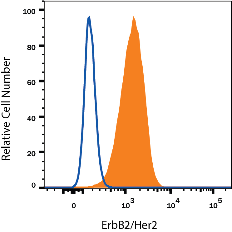
Supportive validation
- Submitted by
- R&D Systems (provider)
- Main image

- Experimental details
- Detection of ErbB2/Her2 in MCF-7 Human Cell Line by Flow Cytometry. MCF-7 human breast cancer cell line was stained with Mouse Anti-Human ErbB2/Her2 Monoclonal Antibody (Catalog # MAB1129, filled histogram) or isotype control antibody (Catalog # MAB0041, open histogram), followed by Phycoerythrin-conjugated Anti-Mouse IgG Secondary Antibody (Catalog # F0102B). View our protocol for Staining Membrane-associated Proteins.