 Explore
 Validate
 Learn
Learn Western blot
Antibody data
- Antibody Data
- Antigen structure
- References [3]
- Comments [0]
- Validations
- Western blot [3]
- Immunocytochemistry [1]
- Immunohistochemistry [1]
- Other assay [2]
Submit
Validation data
Reference
Comment
Report error
- Product number
- PA1-38514 - Provider product page

- Provider
- Invitrogen Antibodies
- Product name
- PSA Polyclonal Antibody
- Antibody type
- Polyclonal
- Antigen
- Purifed from natural sources
- Description
- A suggested positive control is normal prostate or prostate carcinoma.
- Reactivity
- Human
- Host
- Rabbit
- Isotype
- IgG
- Vial size
- 1 mL
- Concentration
- 8.75 mg/mL
- Storage
- -20° C, Avoid Freeze/Thaw Cycles
Submitted references Presence of PSA auto-antibodies in men with prostate abnormalities (prostate cancer/benign prostatic hyperplasia/prostatitis).
Antibodies to prostate-specific antigen in immunoinfertile women and men.
Immunodevice for simultaneous detection of two relevant tumor markers based on separation of different microparticles by dielectrophoresis.
Lokant MT, Naz RK
Andrologia 2015 Apr;47(3):328-32
Andrologia 2015 Apr;47(3):328-32
Antibodies to prostate-specific antigen in immunoinfertile women and men.
Naz RK, Butler TS
Journal of reproductive immunology 2013 Apr;97(2):217-22
Journal of reproductive immunology 2013 Apr;97(2):217-22
Immunodevice for simultaneous detection of two relevant tumor markers based on separation of different microparticles by dielectrophoresis.
Ramón-Azcón J, Yasukawa T, Mizutani F
Biosensors & bioelectronics 2011 Oct 15;28(1):443-9
Biosensors & bioelectronics 2011 Oct 15;28(1):443-9
No comments: Submit comment
Supportive validation
- Submitted by
- Invitrogen Antibodies (provider)
- Main image
- Experimental details
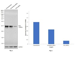
- Knockdown of PSA was achieved by transfecting LNCaP with PSA specific siRNAs (Silencer® select Product # s231157, s200412). Western blot analysis (Fig. a) was performed using whole cell extracts from the PSA knockdown cells (lane 3), non-targeting scrambled siRNA transfected cells (lane 2) and untransfected cells (lane 1). The blot was probed with PSA Polyclonal Antibody (Product # PA1-38514, 1:1000 dilution) and Goat anti-Rabbit IgG (H+L) Superclonal™ Recombinant Secondary Antibody, HRP (Product # A27036, 1:4000 dilution). Densitometric analysis of this western blot is shown in histogram (Fig. b). Decrease in signal upon siRNA mediated knock down confirms that antibody is specific to PSA.

- Submitted by
- Invitrogen Antibodies (provider)
- Main image
- Experimental details
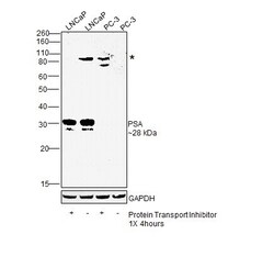
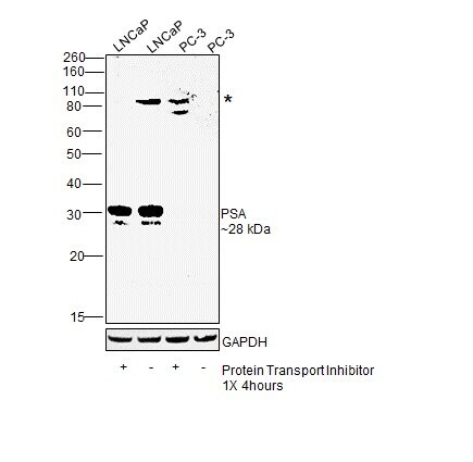
- Western blot was performed using Anti-PSA Polyclonal Antibody(Product # PA1-38514) and a 28kDa band corresponding to PSA, along with an uncharacterized band (*), was observed across positive cell line tested. Whole cell extracts (30 µg lysate) of LNCaP treated with Protein Transport Inhibitor (1X for 4 hours) (Lane 1), LNCaP (Lane 2), PC-3 treated with Protein Transport Inhibitor (1X for 4 hours) (Lane 3) and PC-3 (Lane 4) were electrophoresed using NuPAGE™ 12% Bis-Tris Protein Gel (Product # NP0341BOX). Resolved proteins were then transferred onto a Nitrocellulose membrane (Product # IB23001) by iBlot® 2 Dry Blotting System (Product # IB21001). The blot was probed with the primary antibody (1:1000 dilution) and detected by chemiluminescence with Goat anti-Rabbit IgG (H+L) Superclonal™ Recombinant Secondary Antibody, HRP (Product # A27036, 1:4000 dilution) using the iBright FL 1000 (Product # A32752). Chemiluminescent detection was performed using SuperSignal™ West Dura Extended Duration Substrate (Product # 34076).
- Submitted by
- Invitrogen Antibodies (provider)
- Main image
- Experimental details
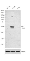
- Western blot was performed using Anti-PSA Polyclonal Antibody (Product # PA1-38514) and a 29kDa band corresponding to PSA was observed in LNCaP but not in DU 145 and PC-3 which are androgen-independent prostate cancer cell lines and are known to have low PSA expression (PubMed: 10232600). Whole cell extracts (30 µg lysate) of DU 145 (Lane 1), LNCaP (Lane 2) and PC-3 (Lane 3) were electrophoresed using NuPAGE™ 4-12% Bis-Tris Protein Gel (Product # NP0321BOX). Resolved proteins were then transferred onto a Nitrocellulose membrane (Product # IB23001) by iBlot® 2 Dry Blotting System (Product # IB21001). The blot was probed with the primary antibody (1:1000 dilution) and detected by chemiluminescence with Goat anti-Rabbit IgG (H+L) Superclonal™ Recombinant Secondary Antibody, HRP (Product # A27036, 1:4000 dilution) using the iBright FL 1000 (Product # A32752). Chemiluminescent detection was performed using Novex® ECL Reagent Kit (Product # WP20005).

Supportive validation
- Submitted by
- Invitrogen Antibodies (provider)
- Main image

- Experimental details
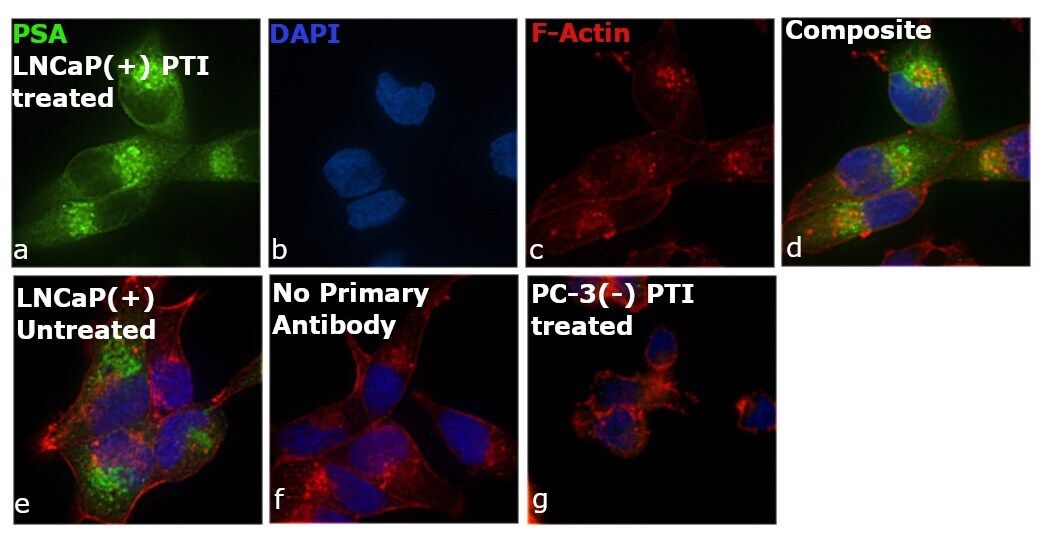
- Immunofluorescence analysis of PSA was performed using 70% confluent log phase LNCaP and PC-3 cells treated with Protein transport inhibitor (1X for 4 hours). The cells were fixed with 4% paraformaldehyde for 10 minutes, permeabilized with 0.1% Triton™ X-100 for 15 minutes, and blocked with 2% BSA for 45 minutes at room temperature. The cells were labeled with PSA Polyclonal Antibody (Product # PA1-38514) at 1:100 dilution in 0.1% BSA, incubated at 4 degree celsius overnight and then labeled with Goat anti-Rabbit IgG (H+L) Highly Cross-Adsorbed Secondary Antibody, Alexa Fluor Plus 488 (Product # A32731), (1:2000 dilution), for 45 minutes at room temperature (Panel a: Green). Nuclei (Panel b:Blue) were stained with ProLong™ Diamond Antifade Mountant with DAPI (Product # P36962). F-actin (Panel c: Red) was stained with Rhodamine Phalloidin (Product # R415, 1:300 dilution). Panel d represents the merged image showing cytoplasmic localization. Panel e represents untreated LNCaP cells. Panel f represents control cells with no primary antibody to assess background. Panel g represents PSA-negative PC-3 cell line showing no staining. The images were captured at 60X magnification.
Supportive validation
- Submitted by
- Invitrogen Antibodies (provider)
- Main image

- Experimental details
- Immunohistochemical analysis of PSA using a polyclonal antibody (Product # PA1-38514).

Supportive validation
- Submitted by
- Invitrogen Antibodies (provider)
- Main image

- Experimental details
- NULL

- Submitted by
- Invitrogen Antibodies (provider)
- Main image

- Experimental details
- NULL
