 Explore
 Validate
 Learn
Learn Western blot
 ELISA
ELISAAntibody data
- Antibody Data
- Antigen structure
- References [1]
- Comments [0]
- Validations
- Western blot [2]
Submit
Validation data
Reference
Comment
Report error
- Product number
- NB600-887 - Provider product page

- Provider
- Novus Biologicals
- Proper citation
- Novus Cat#NB600-887, RRID:AB_2226974
- Product name
- Rabbit Polyclonal Apc10 Antibody
- Antibody type
- Polyclonal
- Description
- Immunogen affinity purified. This detects a band at about 26kD that corresponds to Apc10.
- Reactivity
- Human, Mouse
- Host
- Rabbit
- Isotype
- IgG
- Vial size
- 0.1 mg
- Concentration
- 1.5 mg/ml
- Storage
- Store at -20C. Avoid freeze-thaw cycles.
Submitted references Ube2s regulates Sox2 stability and mouse ES cell maintenance.
Wang J, Zhang Y, Hou J, Qian X, Zhang H, Zhang Z, Li M, Wang R, Liao K, Wang Y, Li Z, Zhong D, Wan P, Dong L, Liu F, Wang X, Wan Y, Xiao W, Zhang WW
Cell death and differentiation 2016 Mar;23(3):393-404
Cell death and differentiation 2016 Mar;23(3):393-404
No comments: Submit comment
Supportive validation
- Submitted by
- Novus Biologicals (provider)
- Main image

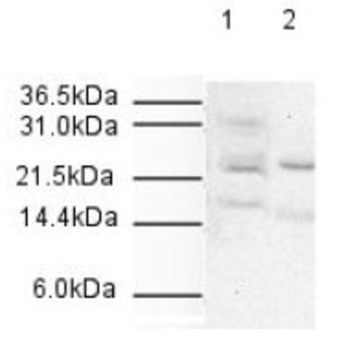
- Experimental details
- Western Blot: Apc10 Antibody [NB600-887] - Antibody was used at a 1:500 dilution to detect human APC10 by western blot. Both HeLa whole cell lysate (lane 1) and nuclear lysate (lane 2) were probed using this antibody. Approximately 20 ug of each lysate was loaded onto a 10% SDS-PAGE gel. Primary antibody was reacted with the membrane at room temperature for 1 h. After subsequent washing, a 1:2,000 dilution of HRP conjugated Gt-a- Rabbit IgG was used for visualization. Exposure time was 5 min. The expected molecular weight of human APC10 is 21 kDa.
- Submitted by
- Novus Biologicals (provider)
- Main image

- Experimental details
- Western Blot: Apc10 Antibody [NB600-887] - Used at a 1:500 dilution to detect human APC10 in various cell extracts. This antibody clearly detects a 26 kDa band corresponding to human APC10 (predicted molecular weight is 21 kDa). All lanes contain 20 ug of lysate or extract as follows: lane 1, HeLa nuclear extract; lane 2, HeLa whole cell lysate; lane 3, A431 whole cell lysate; lane 4, Jurkat whole cell lysate; lane 5, 293 whole cell lysate. Primary antibody was reacted with the membrane at room temperature for 1 h. After subsequent washing, a 1:5,000 dilution of HRP conjugated Gt-a-Rabbit IgG was used for visualization. Exposure time was 4 min.
