 Explore
 Validate
 Learn
Learn Western blot
 Immunohistochemistry
Antibody data
- Antibody Data
- Antigen structure
- References [38]
- Comments [0]
- Validations
- Immunohistochemistry [1]
- Other assay [19]
Submit
Validation data
Reference
Comment
Report error
- Product number
- 35-6200 - Provider product page

- Provider
- Invitrogen Antibodies
- Product name
- NY-ESO-1 Monoclonal Antibody (E978)
- Antibody type
- Monoclonal
- Antigen
- Recombinant full-length protein
- Reactivity
- Human
- Host
- Mouse
- Isotype
- IgG
- Antibody clone number
- E978
- Vial size
- 100 µg
- Concentration
- 0.5 mg/mL
- Storage
- Store at 4°C short term. For long term storage, store at -20°C, avoiding freeze/thaw cycles.
Submitted references Myeloid-derived suppressor cells infiltration in non-small-cell lung cancer tumor and MAGE-A4 and NY-ESO-1 expression.
Sublethal Radiation Affects Antigen Processing and Presentation Genes to Enhance Immunogenicity of Cancer Cells.
Extramammary Paget disease shows differential expression of B7 family members B7-H3, B7-H4, PD-L1, PD-L2 and cancer/testis antigens NY-ESO-1 and MAGE-A.
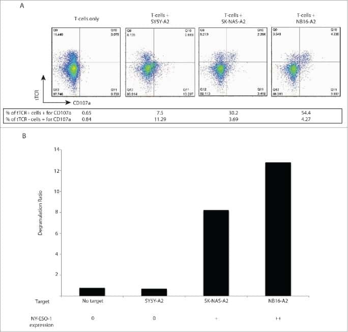
Immune landscape and in vivo immunogenicity of NY-ESO-1 tumor antigen in advanced neuroblastoma patients.
Expression and immunogenicity of NY-ESO-1 in colorectal cancer.
T cells targeting NY-ESO-1 demonstrate efficacy against disseminated neuroblastoma.
Induction of cancer testis antigen expression in circulating acute myeloid leukemia blasts following hypomethylating agent monotherapy.
Treatment with 5-Aza-2'-Deoxycytidine Induces Expression of NY-ESO-1 and Facilitates Cytotoxic T Lymphocyte-Mediated Tumor Cell Killing.
NY-ESO-1 (CTAG1B) expression in mesenchymal tumors.
Immunomodulatory action of SGI-110, a hypomethylating agent, in acute myeloid leukemia cells and xenografts.
Decitabine facilitates immune recognition of sarcoma cells by upregulating CT antigens, MHC molecules, and ICAM-1.
Induction of antigen-specific immunity with a vaccine targeting NY-ESO-1 to the dendritic cell receptor DEC-205.
Gene expression profiling using nanostring digital RNA counting to identify potential target antigens for melanoma immunotherapy.
Expression of MAGE-C1/CT7 and selected cancer/testis antigens in ovarian borderline tumours and primary and recurrent ovarian carcinomas.
Potential therapeutic value of dendritic cells loaded with NY‑ESO‑1 protein for the immunotherapy of advanced hepatocellular carcinoma.
Effector memory and central memory NY-ESO-1-specific re-directed T cells for treatment of multiple myeloma.
NY-ESO-1 expression in hepatocellular carcinoma: A potential new marker for early recurrence after surgery.
Ex vivo enrichment of circulating anti-tumor T cells from both cutaneous and ocular melanoma patients: clinical implications for adoptive cell transfer therapy.
MAGE-C2/CT10 protein expression is an independent predictor of recurrence in prostate cancer.
Antibody responses to NY-ESO-1 in primary breast cancer identify a subtype target for immunotherapy.
γ-Radiation promotes immunological recognition of cancer cells through increased expression of cancer-testis antigens in vitro and in vivo.
MAGE-A1, MAGE-A3, and NY-ESO-1 can be upregulated on neuroblastoma cells to facilitate cytotoxic T lymphocyte-mediated tumor cell killing.
Fucoidin enhances dendritic cell-mediated T-cell cytotoxicity against NY-ESO-1 expressing human cancer cells.
Immunobiological characterization of cancer stem cells isolated from glioblastoma patients.
Modified tumour antigen-encoding mRNA facilitates the analysis of naturally occurring and vaccine-induced CD4 and CD8 T cells in cancer patients.
p53 autoantibodies, cytokine levels and ovarian carcinogenesis.
Frequent expression of the novel cancer testis antigen MAGE-C2/CT-10 in hepatocellular carcinoma.
Tumor-infiltrating T cells correlate with NY-ESO-1-specific autoantibodies in ovarian cancer.
MAGE-C1/CT-7 expression in plasma cell myeloma: sub-cellular localization impacts on clinical outcome.
MAD-CT-2 identified as a novel melanoma cancer-testis antigen using phage immunoblot analysis.
NY-ESO-1 protein expression in primary breast carcinoma and metastases: correlation with CD8+ T-cell and CD79a+ plasmacytic/B-cell infiltration.
Oral premalignant lesions induce immune reactivity to both premalignant oral lesions and head and neck squamous cell carcinoma.
Cancer-germline gene expression in pediatric solid tumors using quantitative real-time PCR.
Restriction of GAGE protein expression to subpopulations of cancer cells is independent of genotype and may limit the use of GAGE proteins as targets for cancer immunotherapy.
Quantifying and imaging NY-ESO-1/LAGE-1-derived epitopes on tumor cells using high affinity T cell receptors.
Heterogeneous expression of GAGE, NY-ESO-1, MAGE-A and SSX proteins in esophageal cancer: Implications for immunotherapy.
Tumor infiltrating lymphocytes in seminoma lesions comprise clonally expanded cytotoxic T cells.
Expression profile of cancer-testis genes in 121 human colorectal cancer tissue and adjacent normal tissue.
Hou Z, Liang X, Wang X, Zhou Z, Shi G
Oncology letters 2020 Jun;19(6):3982-3992
Oncology letters 2020 Jun;19(6):3982-3992
Sublethal Radiation Affects Antigen Processing and Presentation Genes to Enhance Immunogenicity of Cancer Cells.
Punnanitinont A, Kannisto ED, Matsuzaki J, Odunsi K, Yendamuri S, Singh AK, Patnaik SK
International journal of molecular sciences 2020 Apr 7;21(7)
International journal of molecular sciences 2020 Apr 7;21(7)
Extramammary Paget disease shows differential expression of B7 family members B7-H3, B7-H4, PD-L1, PD-L2 and cancer/testis antigens NY-ESO-1 and MAGE-A.
Pourmaleki M, Young JH, Socci ND, Chiang S, Edelweiss M, Li Y, Zhang M, Roshal L, Chi DS, Busam KJ, Mellinghoff IK, Hollmann TJ
Oncotarget 2019 Oct 22;10(58):6152-6167
Oncotarget 2019 Oct 22;10(58):6152-6167
Immune landscape and in vivo immunogenicity of NY-ESO-1 tumor antigen in advanced neuroblastoma patients.
Camisaschi C, Renne SL, Beretta V, Rini F, Spagnuolo RD, Tuccitto A, Podda MG, Parmiani G, Rivoltini L, Collini P, Castelli C, Luksch R
BMC cancer 2018 Oct 16;18(1):983
BMC cancer 2018 Oct 16;18(1):983
Expression and immunogenicity of NY-ESO-1 in colorectal cancer.
Li Y, Song R, Li X, Xu F
Experimental and therapeutic medicine 2017 Jun;13(6):3581-3585
Experimental and therapeutic medicine 2017 Jun;13(6):3581-3585
T cells targeting NY-ESO-1 demonstrate efficacy against disseminated neuroblastoma.
Singh N, Kulikovskaya I, Barrett DM, Binder-Scholl G, Jakobsen B, Martinez D, Pawel B, June CH, Kalos MD, Grupp SA
Oncoimmunology 2016;5(1):e1040216
Oncoimmunology 2016;5(1):e1040216
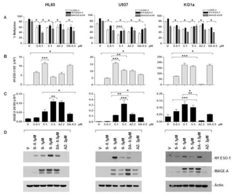
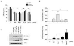
Induction of cancer testis antigen expression in circulating acute myeloid leukemia blasts following hypomethylating agent monotherapy.
Srivastava P, Paluch BE, Matsuzaki J, James SR, Collamat-Lai G, Blagitko-Dorfs N, Ford LA, Naqash R, Lübbert M, Karpf AR, Nemeth MJ, Griffiths EA
Oncotarget 2016 Mar 15;7(11):12840-56
Oncotarget 2016 Mar 15;7(11):12840-56
Treatment with 5-Aza-2'-Deoxycytidine Induces Expression of NY-ESO-1 and Facilitates Cytotoxic T Lymphocyte-Mediated Tumor Cell Killing.
Klar AS, Gopinadh J, Kleber S, Wadle A, Renner C
PloS one 2015;10(10):e0139221
PloS one 2015;10(10):e0139221
NY-ESO-1 (CTAG1B) expression in mesenchymal tumors.
Endo M, de Graaff MA, Ingram DR, Lim S, Lev DC, Briaire-de Bruijn IH, Somaiah N, Bovée JV, Lazar AJ, Nielsen TO
Modern pathology : an official journal of the United States and Canadian Academy of Pathology, Inc 2015 Apr;28(4):587-95
Modern pathology : an official journal of the United States and Canadian Academy of Pathology, Inc 2015 Apr;28(4):587-95
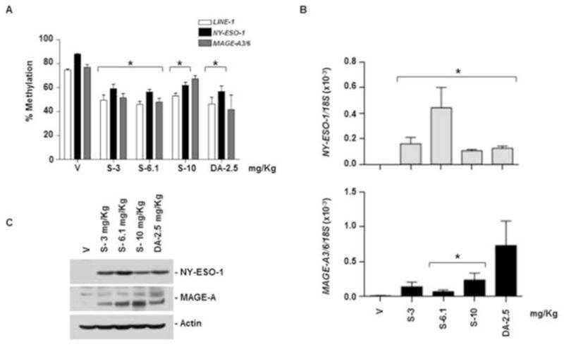
Immunomodulatory action of SGI-110, a hypomethylating agent, in acute myeloid leukemia cells and xenografts.
Srivastava P, Paluch BE, Matsuzaki J, James SR, Collamat-Lai G, Karbach J, Nemeth MJ, Taverna P, Karpf AR, Griffiths EA
Leukemia research 2014 Nov;38(11):1332-41
Leukemia research 2014 Nov;38(11):1332-41
Decitabine facilitates immune recognition of sarcoma cells by upregulating CT antigens, MHC molecules, and ICAM-1.
Krishnadas DK, Bao L, Bai F, Chencheri SC, Lucas K
Tumour biology : the journal of the International Society for Oncodevelopmental Biology and Medicine 2014 Jun;35(6):5753-62
Tumour biology : the journal of the International Society for Oncodevelopmental Biology and Medicine 2014 Jun;35(6):5753-62
Induction of antigen-specific immunity with a vaccine targeting NY-ESO-1 to the dendritic cell receptor DEC-205.
Dhodapkar MV, Sznol M, Zhao B, Wang D, Carvajal RD, Keohan ML, Chuang E, Sanborn RE, Lutzky J, Powderly J, Kluger H, Tejwani S, Green J, Ramakrishna V, Crocker A, Vitale L, Yellin M, Davis T, Keler T
Science translational medicine 2014 Apr 16;6(232):232ra51
Science translational medicine 2014 Apr 16;6(232):232ra51
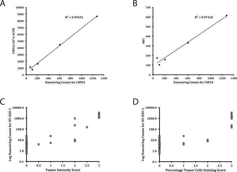
Gene expression profiling using nanostring digital RNA counting to identify potential target antigens for melanoma immunotherapy.
Beard RE, Abate-Daga D, Rosati SF, Zheng Z, Wunderlich JR, Rosenberg SA, Morgan RA
Clinical cancer research : an official journal of the American Association for Cancer Research 2013 Sep 15;19(18):4941-50
Clinical cancer research : an official journal of the American Association for Cancer Research 2013 Sep 15;19(18):4941-50
Expression of MAGE-C1/CT7 and selected cancer/testis antigens in ovarian borderline tumours and primary and recurrent ovarian carcinomas.
Zimmermann AK, Imig J, Klar A, Renner C, Korol D, Fink D, Stadlmann S, Singer G, Knuth A, Moch H, Caduff R
Virchows Archiv : an international journal of pathology 2013 May;462(5):565-74
Virchows Archiv : an international journal of pathology 2013 May;462(5):565-74
Potential therapeutic value of dendritic cells loaded with NY‑ESO‑1 protein for the immunotherapy of advanced hepatocellular carcinoma.
Chen Y, Huang A, Gao M, Yan Y, Zhang W
International journal of molecular medicine 2013 Dec;32(6):1366-72
International journal of molecular medicine 2013 Dec;32(6):1366-72
Effector memory and central memory NY-ESO-1-specific re-directed T cells for treatment of multiple myeloma.
Schuberth PC, Jakka G, Jensen SM, Wadle A, Gautschi F, Haley D, Haile S, Mischo A, Held G, Thiel M, Tinguely M, Bifulco CB, Fox BA, Renner C, Petrausch U
Gene therapy 2013 Apr;20(4):386-95
Gene therapy 2013 Apr;20(4):386-95
NY-ESO-1 expression in hepatocellular carcinoma: A potential new marker for early recurrence after surgery.
Xu H, Gu N, Liu ZB, Zheng M, Xiong F, Wang SY, Li N, Lu J
Oncology letters 2012 Jan;3(1):39-44
Oncology letters 2012 Jan;3(1):39-44
Ex vivo enrichment of circulating anti-tumor T cells from both cutaneous and ocular melanoma patients: clinical implications for adoptive cell transfer therapy.
Mazzarella T, Cambiaghi V, Rizzo N, Pilla L, Parolini D, Orsenigo E, Colucci A, Modorati G, Doglioni C, Parmiani G, Maccalli C
Cancer immunology, immunotherapy : CII 2012 Aug;61(8):1169-82
Cancer immunology, immunotherapy : CII 2012 Aug;61(8):1169-82
MAGE-C2/CT10 protein expression is an independent predictor of recurrence in prostate cancer.
von Boehmer L, Keller L, Mortezavi A, Provenzano M, Sais G, Hermanns T, Sulser T, Jungbluth AA, Old LJ, Kristiansen G, van den Broek M, Moch H, Knuth A, Wild PJ
PloS one 2011;6(7):e21366
PloS one 2011;6(7):e21366
Antibody responses to NY-ESO-1 in primary breast cancer identify a subtype target for immunotherapy.
Hamaï A, Duperrier-Amouriaux K, Pignon P, Raimbaud I, Memeo L, Colarossi C, Canzonieri V, Perin T, Classe JM, Campone M, Jézéquel P, Campion L, Ayyoub M, Valmori D
PloS one 2011;6(6):e21129
PloS one 2011;6(6):e21129
γ-Radiation promotes immunological recognition of cancer cells through increased expression of cancer-testis antigens in vitro and in vivo.
Sharma A, Bode B, Wenger RH, Lehmann K, Sartori AA, Moch H, Knuth A, Boehmer Lv, Broek Mv
PloS one 2011;6(11):e28217
PloS one 2011;6(11):e28217
MAGE-A1, MAGE-A3, and NY-ESO-1 can be upregulated on neuroblastoma cells to facilitate cytotoxic T lymphocyte-mediated tumor cell killing.
Bao L, Dunham K, Lucas K
Cancer immunology, immunotherapy : CII 2011 Sep;60(9):1299-307
Cancer immunology, immunotherapy : CII 2011 Sep;60(9):1299-307
Fucoidin enhances dendritic cell-mediated T-cell cytotoxicity against NY-ESO-1 expressing human cancer cells.
Hu Y, Cheng SC, Chan KT, Ke Y, Xue B, Sin FW, Zeng C, Xie Y
Biochemical and biophysical research communications 2010 Feb 12;392(3):329-34
Biochemical and biophysical research communications 2010 Feb 12;392(3):329-34
Immunobiological characterization of cancer stem cells isolated from glioblastoma patients.
Di Tomaso T, Mazzoleni S, Wang E, Sovena G, Clavenna D, Franzin A, Mortini P, Ferrone S, Doglioni C, Marincola FM, Galli R, Parmiani G, Maccalli C
Clinical cancer research : an official journal of the American Association for Cancer Research 2010 Feb 1;16(3):800-13
Clinical cancer research : an official journal of the American Association for Cancer Research 2010 Feb 1;16(3):800-13
Modified tumour antigen-encoding mRNA facilitates the analysis of naturally occurring and vaccine-induced CD4 and CD8 T cells in cancer patients.
Knights AJ, Nuber N, Thomson CW, de la Rosa O, Jäger E, Tiercy JM, van den Broek M, Pascolo S, Knuth A, Zippelius A
Cancer immunology, immunotherapy : CII 2009 Mar;58(3):325-38
Cancer immunology, immunotherapy : CII 2009 Mar;58(3):325-38
p53 autoantibodies, cytokine levels and ovarian carcinogenesis.
Tsai-Turton M, Santillan A, Lu D, Bristow RE, Chan KC, Shih IeM, Roden RB
Gynecologic oncology 2009 Jul;114(1):12-7
Gynecologic oncology 2009 Jul;114(1):12-7
Frequent expression of the novel cancer testis antigen MAGE-C2/CT-10 in hepatocellular carcinoma.
Riener MO, Wild PJ, Soll C, Knuth A, Jin B, Jungbluth A, Hellerbrand C, Clavien PA, Moch H, Jochum W
International journal of cancer 2009 Jan 15;124(2):352-7
International journal of cancer 2009 Jan 15;124(2):352-7
Tumor-infiltrating T cells correlate with NY-ESO-1-specific autoantibodies in ovarian cancer.
Milne K, Barnes RO, Girardin A, Mawer MA, Nesslinger NJ, Ng A, Nielsen JS, Sahota R, Tran E, Webb JR, Wong MQ, Wick DA, Wray A, McMurtrie E, Köbel M, Kalloger SE, Gilks CB, Watson PH, Nelson BH
PloS one 2008;3(10):e3409
PloS one 2008;3(10):e3409
MAGE-C1/CT-7 expression in plasma cell myeloma: sub-cellular localization impacts on clinical outcome.
Tinguely M, Jenni B, Knights A, Lopes B, Korol D, Rousson V, Curioni Fontecedro A, Cogliatti SB, Bittermann AG, Schmid U, Dommann-Scherrer C, Maurer R, Renner C, Probst-Hensch NM, Moch H, Knuth A, Zippelius A
Cancer science 2008 Apr;99(4):720-5
Cancer science 2008 Apr;99(4):720-5
MAD-CT-2 identified as a novel melanoma cancer-testis antigen using phage immunoblot analysis.
Dubovsky JA, Albertini MR, McNeel DG
Journal of immunotherapy (Hagerstown, Md. : 1997) 2007 Oct;30(7):675-83
Journal of immunotherapy (Hagerstown, Md. : 1997) 2007 Oct;30(7):675-83
NY-ESO-1 protein expression in primary breast carcinoma and metastases: correlation with CD8+ T-cell and CD79a+ plasmacytic/B-cell infiltration.
Theurillat JP, Ingold F, Frei C, Zippelius A, Varga Z, Seifert B, Chen YT, Jäger D, Knuth A, Moch H
International journal of cancer 2007 Jun 1;120(11):2411-7
International journal of cancer 2007 Jun 1;120(11):2411-7
Oral premalignant lesions induce immune reactivity to both premalignant oral lesions and head and neck squamous cell carcinoma.
Young MR, Neville BW, Chi AC, Lathers DM, Boyd Gillespie M, Day TA
Cancer immunology, immunotherapy : CII 2007 Jul;56(7):1077-86
Cancer immunology, immunotherapy : CII 2007 Jul;56(7):1077-86
Cancer-germline gene expression in pediatric solid tumors using quantitative real-time PCR.
Jacobs JF, Brasseur F, Hulsbergen-van de Kaa CA, van de Rakt MW, Figdor CG, Adema GJ, Hoogerbrugge PM, Coulie PG, de Vries IJ
International journal of cancer 2007 Jan 1;120(1):67-74
International journal of cancer 2007 Jan 1;120(1):67-74
Restriction of GAGE protein expression to subpopulations of cancer cells is independent of genotype and may limit the use of GAGE proteins as targets for cancer immunotherapy.
Gjerstorff MF, Johansen LE, Nielsen O, Kock K, Ditzel HJ
British journal of cancer 2006 Jun 19;94(12):1864-73
British journal of cancer 2006 Jun 19;94(12):1864-73
Quantifying and imaging NY-ESO-1/LAGE-1-derived epitopes on tumor cells using high affinity T cell receptors.
Purbhoo MA, Sutton DH, Brewer JE, Mullings RE, Hill ME, Mahon TM, Karbach J, Jäger E, Cameron BJ, Lissin N, Vyas P, Chen JL, Cerundolo V, Jakobsen BK
Journal of immunology (Baltimore, Md. : 1950) 2006 Jun 15;176(12):7308-16
Journal of immunology (Baltimore, Md. : 1950) 2006 Jun 15;176(12):7308-16
Heterogeneous expression of GAGE, NY-ESO-1, MAGE-A and SSX proteins in esophageal cancer: Implications for immunotherapy.
Akcakanat A, Kanda T, Tanabe T, Komukai S, Yajima K, Nakagawa S, Ohashi M, Hatakeyama K
International journal of cancer 2006 Jan 1;118(1):123-8
International journal of cancer 2006 Jan 1;118(1):123-8
Tumor infiltrating lymphocytes in seminoma lesions comprise clonally expanded cytotoxic T cells.
Hadrup SR, Braendstrup O, Jacobsen GK, Mortensen S, Pedersen LØ, Seremet T, Andersen MH, Becker JC, Straten PT
International journal of cancer 2006 Aug 15;119(4):831-8
International journal of cancer 2006 Aug 15;119(4):831-8
Expression profile of cancer-testis genes in 121 human colorectal cancer tissue and adjacent normal tissue.
Li M, Yuan YH, Han Y, Liu YX, Yan L, Wang Y, Gu J
Clinical cancer research : an official journal of the American Association for Cancer Research 2005 Mar 1;11(5):1809-14
Clinical cancer research : an official journal of the American Association for Cancer Research 2005 Mar 1;11(5):1809-14
No comments: Submit comment
Supportive validation
- Submitted by
- Invitrogen Antibodies (provider)
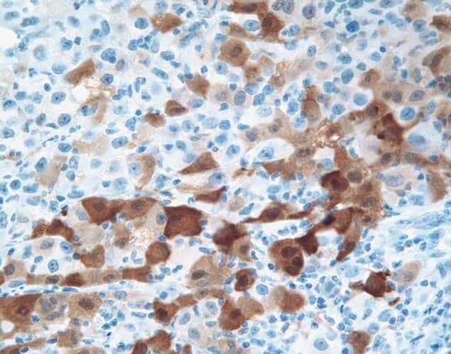
- Main image

- Experimental details
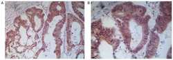
- Immunohistochemical staining of melanoma tissue using mouse anti-NY-ESO-1 monoclonal antibody (clone E978) (Product # 35-6200).
Supportive validation
- Submitted by
- Invitrogen Antibodies (provider)
- Main image
- Experimental details
- NULL
- Submitted by
- Invitrogen Antibodies (provider)
- Main image

- Experimental details
- NULL
- Submitted by
- Invitrogen Antibodies (provider)
- Main image

- Experimental details
- NULL
- Submitted by
- Invitrogen Antibodies (provider)
- Main image

- Experimental details
- NULL
- Submitted by
- Invitrogen Antibodies (provider)
- Main image
- Experimental details
- NULL
- Submitted by
- Invitrogen Antibodies (provider)
- Main image

- Experimental details
- NULL
- Submitted by
- Invitrogen Antibodies (provider)
- Main image

- Experimental details
- NULL
- Submitted by
- Invitrogen Antibodies (provider)
- Main image
- Experimental details
- NULL
- Submitted by
- Invitrogen Antibodies (provider)
- Main image
- Experimental details
- NULL
- Submitted by
- Invitrogen Antibodies (provider)
- Main image

- Experimental details
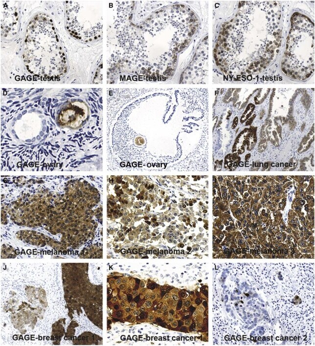
- Figure 2 Comparison of GAGE, MAGE-1 and NY-ESO-1 staining in normal testis, and analysis of GAGE protein expression in ovary and different types of cancer. GAGE ( A ), MAGE ( B ) and NY-ESO-1 ( C ) were detected in spermatogonia and primary spermatocytes of the seminiferous tubuli. However, while MAGE-A1 and NY-ESO-1 were located only in the cytoplasm of these cells, GAGE staining was also present, and more intense, in the nuclei. GAGE was also expressed in oocytes of resting ( D ) and maturing ( E ) follicles of normal ovaries, which also contained GAGE-negative oocytes. In three malignant melanomas, all cells exhibited cytoplasmic staining ( G - I ), whereas nuclear staining was observed in only two melanomas ( G and H ). Heterogeneous staining was also seen in other types of cancer, including lung carcinoma ( F ) and breast carcinoma ( J - L ). One breast carcinoma exhibited variations in both cytoplasmic and nuclear GAGE staining among cells ( J and K ), while only few cells of another breast cancer specimen were positive ( L ). Magnification: x 10 ( A , B , C , E , F , J , L ), x 20 ( G - I ), x 40 ( D , K ).
- Submitted by
- Invitrogen Antibodies (provider)
- Main image
- Experimental details
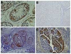
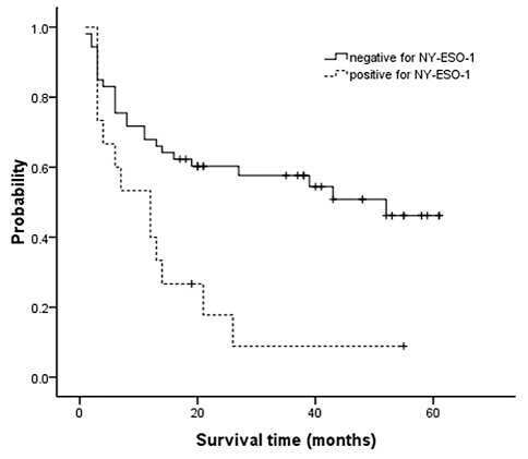
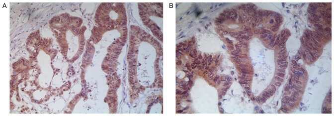
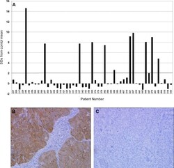
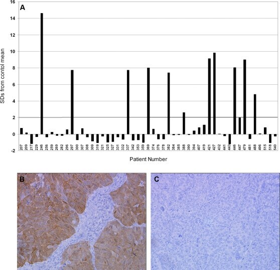
- Figure 3 NY-ESO-1 serum autoantibodies and antigen expression in high-grade serous ovarian cancer. (A) Serum autoantibody responses to NY-ESO-1 in the 35-patient cohort. Autoantibody responses are reported as the number of standard deviations from the mean of 60 age- and gender-matched controls with no known personal history of cancer. (B,C) Immunohistochemical analysis of NY-ESO-1 expression in two representative serous ovarian tumors with high (B) and negative (C) expression of the antigen.
- Submitted by
- Invitrogen Antibodies (provider)
- Main image

- Experimental details
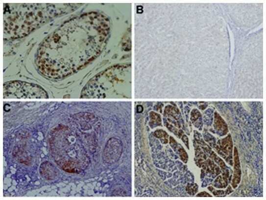
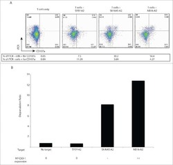
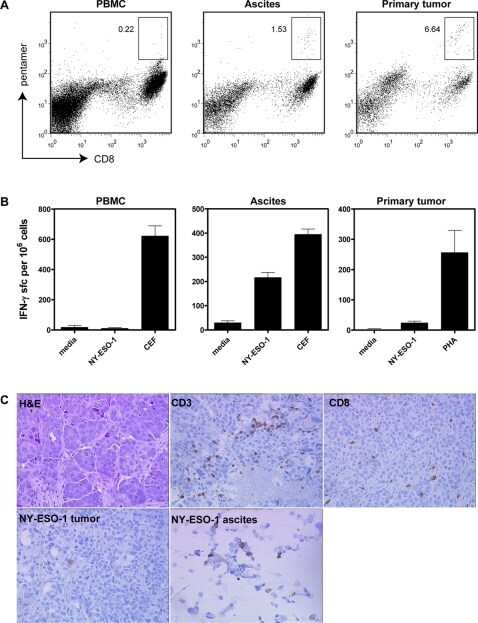
- Figure 4 Analysis of the T cell response in a patient with autoantibodies to NY-ESO-1 showing the presence of NY-ESO-1 reactive CD8+ T cells in ascites and tumor despite the lack of NY-ESO-1 expression in solid tumor. (A) MHC class I pentamer analysis demonstrating enrichment of NY-ESO-1-specific CD8+ T cells in ascites and solid tumor compared to peripheral blood. The boxed areas and associated numbers represent the percentage of pentamer-positive cells relative to total CD8+ cells. (B) ELISPOT analysis of IFN-gamma production by T cells after stimulation with an HLA-A2-binding peptide from NY-ESO-1. Data is presented as the number of IFN-gamma-producing cells per 1x10 6 bulk cells from the indicated tissue compartments. (C) Immunohistochemical analysis of tumor-infiltrating CD3+ and CD8+ T cells in tumor stroma, and expression of NY-ESO-1 antigen. While the solid tumor was negative for expression of NY-ESO-1, a fraction of cells from ascites were positive. The cellular fraction of ascites also contained cytokeratin-positive epithelial cells, presumably of tumor origin (data not shown).
- Submitted by
- Invitrogen Antibodies (provider)
- Main image
- Experimental details
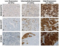
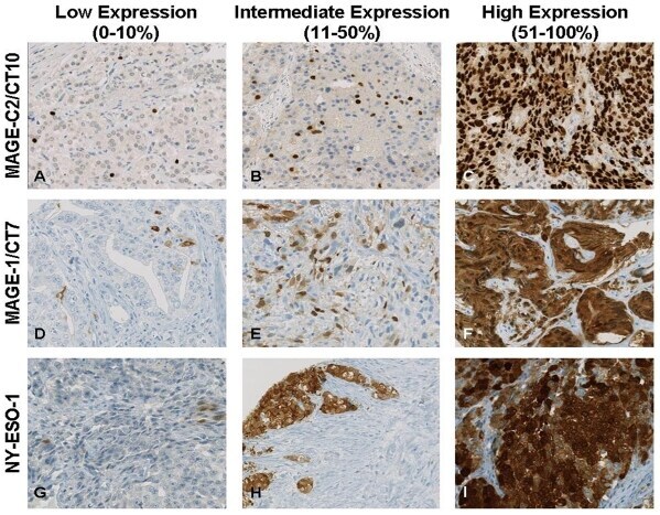
- Figure 2 Representative immunohistochemical expression patterns from 1-10%, 11-50% and 51-100% of MAGE-C2/CT10 (A, B and C), MAGE-C1/CT7 (D, E and F) and NY-ESO-1 (G, H and I). A: Radical prostatectomy specimen (Gleason 4+3), B: Bone metastasis of prostate cancer, C: Bone metastasis of prostate cancer, D: Radical prostatectomy specimen (Gleason 4+3), E: Castration-resistant prostate cancer (Gleason 5+5), F: Palliative transurethral resection of prostate cancer (Gleason 4+5), G: Bone metastasis of prostate cancer, H: Palliative transurethral resection of prostate cancer (Gleason 5+4), I: Castration-resistant prostate cancer (Gleason 5+4). Original magnification: 200x; Magnification bar: 20 uM.
- Submitted by
- Invitrogen Antibodies (provider)
- Main image
- Experimental details
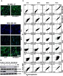
- Figure 2 gamma-radiation up-regulates CT-antigens and MHC-I molecules on the protein level. (A) Immunofluorescence of MDA-MB-469 and SK-MEL-37 cells exposed to a single dose irradiation of 20 Gy and stained with antibodies against NY-ESO-1 and CT7 at different time points after irradiation. (B) MDA-MB-469, SK-MEL-37 and MCF 10A cell lines were exposed to a single dose of 20 Gy and the expression of NY-ESO-1 and CT7 was analyzed by Western blotting 72 h later. (C) breast cancer and (D) osteosarcoma cell lines were exposed to single dose irradiation of 20 Gy and the expression of HLA-ABC and beta 2 microglobulin was quantified at different time points after irradiation by flow cytometry. RAD: indicates gamma-radiation.

- Submitted by
- Invitrogen Antibodies (provider)
- Main image
- Experimental details
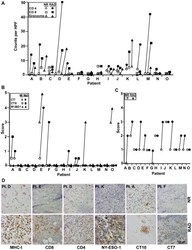
- Figure 5 Radiotherapy induced expression of CT-antigens and MHC-I and lymphocyte infiltration in sarcoma patients. Paraffin-embedded paired tissue sections obtained from sarcoma patients (n = 15) before and after irradiation were analyzed by immunohistochemistry for (A) presence of CD8 + , CD4 + and granzyme + cells, (B) expression of CT7, NY-ESO-1 and CT10 and (C) MHC-I expression. The CT-antigens were scored as the mean percentage of live cells expressing the antigen to the total number of cells in five high power fields (40X objective). The infiltration by lymphocytes was taken as the mean by counting the number of cells expressing CD4, CD8 and granzyme in five high power fields, and MHC-I was scored based on the intensity of the staining. (D) Representative sections showing the expression of CT-antigens and MHC-I and infiltration of lymphocytes before and after radiotherapy by immunohistochemistry. Depicted are: patient F for the expression of CT7, patient A for CT10, patient K for NY-ESO-1, patient D for CD4 and MHC-I and patient E for CD8 expression. NR indicates non-radiated and RAD indicates corresponding irradiated sections. Pt: indicates patient number. Patient information is listed in supplementary Table S2 .

- Submitted by
- Invitrogen Antibodies (provider)
- Main image
- Experimental details
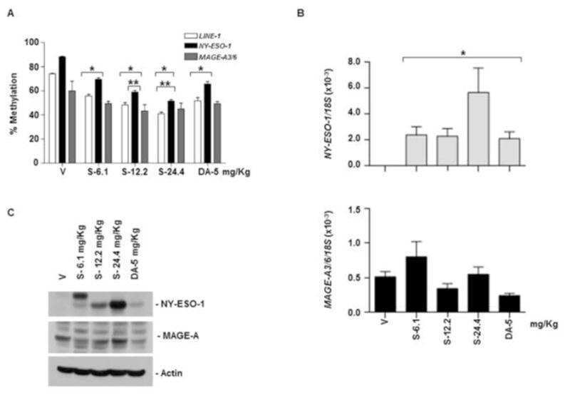
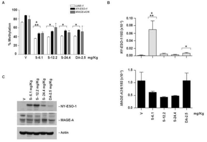
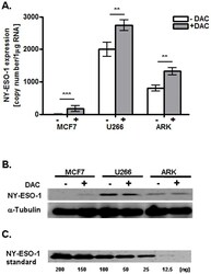
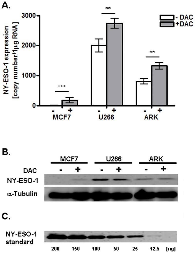
- Fig 2 Effects of DAC treatment on NY-ESO-1 mRNA and protein expression in MCF7, U266, and ARK cell lines. A. NY-ESO-1 specific mRNA, quantified as copy numbers/mug RNA using qRT-PCR. All data are representative of three independent experiments performed in triplicate. B. NY-ESO-1 protein expression analyzed by Western blotting (n = 3). The first and second line show total cell lysate of the respective cell line +/- DAC treatment and detection via a NY-ESO-1 specific or alpha-Tubulin specific (loading control) antibody. The third line shows a Western blot of recombinant NY-ESO-1 protein as control.
- Submitted by
- Invitrogen Antibodies (provider)
- Main image
- Experimental details
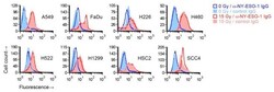
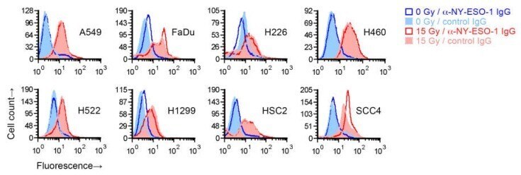
- Figure 2 Effect of radiation on cell surface NY-ESO-1 measurement by fluorescence flow cytometry. Sub-confluent adherent cultures of indicated cell lines that had been grown in parallel to same cell density were treated with 0 or 15 Gy X-rays. After four days, adherent cells were collected by scraping and examined by flow cytometry for binding of an unconjugated mouse IgG1 antibody against NY-ESO-1. Antibody binding was indirectly detected with a fluorophore-conjugated rat antibody against mouse IgG. Binding of a control IgG1 (normal mouse serum fraction) was similarly detected. Shown are representative histograms of viable cells identified by 7 -amino-actinomycin D staining.
- Submitted by
- Invitrogen Antibodies (provider)
- Main image

- Experimental details
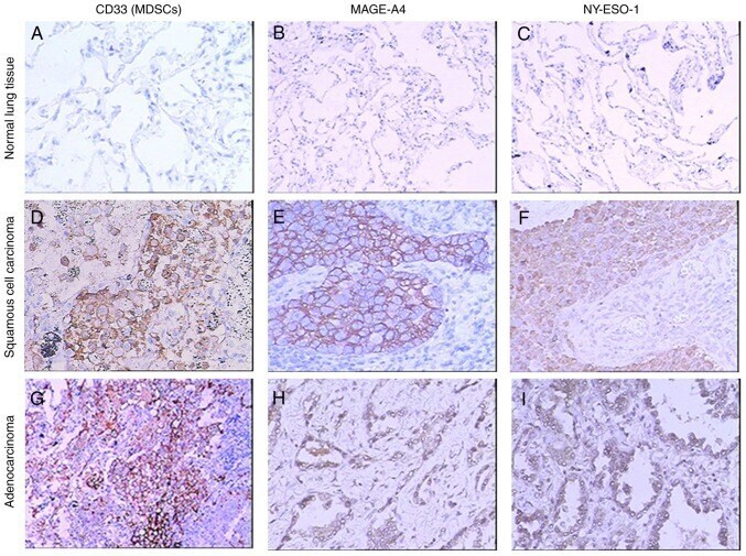
- Figure 1. Immunohistochemical findings for MAGE-A4, NY-ESO-1 and CD33 in non-small cell lung cancer tissues. Specimens from the normal lung were used as a negative control. Staining of the normal lung demonstrated no reactivity for (A) CD33, (B) MAGE-A4 and (C) NY-ESO-1. Expression of (D) CD33, (E) MAGE-A4 and (F) NY-ESO-1 in squamous cell carcinoma. Expression of (G) CD33, (H) MAGE-A4 and (I) NY-ESO-1 in adenocarcinoma. Magnification, x100. MDSCs, myeloid derived suppressor cells; MAGE-A4, melanoma-associated antigen 4; NY-ESO-1, New York esophageal squamous cell carcinoma-1.
- Submitted by
- Invitrogen Antibodies (provider)
- Main image
- Experimental details
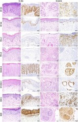
- Figure 1 Representative images of immunohistochemistry for B7-H3, B7-H4, PD-L1, PD-L2, MAGE-A, NY-ESO-1, MHC-I, and B2M in in situ and invasive EMPD. IHC images for each marker along with the corresponding H&E image for each case are shown for in situ and invasive EMPD.
