 Explore
 Validate
 Learn
Learn17-1699-42
antibody from Invitrogen Antibodies
Targeting: SIGLEC1
CD169, dJ1009E24.1, FLJ00051, FLJ00055, FLJ00073, FLJ32150, sialoadhesin, SIGLEC-1, SN
 Flow cytometry
Antibody data
- Antibody Data
- Antigen structure
- References [5]
- Comments [0]
- Validations
- Flow cytometry [1]
- Other assay [4]
Submit
Validation data
Reference
Comment
Report error
- Product number
- 17-1699-42 - Provider product page

- Provider
- Invitrogen Antibodies
- Product name
- CD169 (Siglec-1) Monoclonal Antibody (7-239), APC, eBioscience™
- Antibody type
- Monoclonal
- Antigen
- Other
- Description
- Description: This 7-239 monoclonal antibody reacts with human CD169, also known as Siglec-1 and Sialoadhesin. This type I transmembrane protein is a member of the sialic acid binding Ig-like lectin (Siglec) family. Expressed on macrophages and dendritic cells in peripheral blood, lymph nodes, and spleen in humans, CD169 binds glycoproteins and glycolipids containing N-acetylneuraminyl alpha 2-3-galactose. CD169 expression can also be induced on CD14+ monocytes by interferon-alpha and interferon-gamma to mediate HIV-1 infection. Finally, CD169 has also been shown to be involved in antigen presentation to invariant NK T cells. This monoclonal antibody has been reported to inhibit erythrocyte binding to CD169-expressing cells. Applications Reported: This 7-239 antibody has been reported for use in flow cytometric analysis. Applications Tested: This 7-239 antibody has been pre-titrated and tested by flow cytometric analysis of cultured human peripheral blood cells. This can be used at 5 µL (0.06 µg) per test. A test is defined as the amount (µg) of antibody that will stain a cell sample in a final volume of 100 µL. Cell number should be determined empirically but can range from 10^5 to 10^8 cells/test. Excitation: 633-647 nm; Emission: 660 nm; Laser: Red Laser. Filtration: 0.2 µm post-manufacturing filtered.
- Reactivity
- Human
- Host
- Mouse
- Isotype
- IgG
- Antibody clone number
- 7-239
- Vial size
- 100 Tests
- Concentration
- 5 µL/Test
- Storage
- 4° C, store in dark, DO NOT FREEZE!
Submitted references Decreased expression of a phagocytic receptor Siglec-1 on alveolar macrophages in chronic obstructive pulmonary disease.
Phenotypic and Functional Characterization of Monoclonal Antibodies with Specificity for Rhesus Macaque CD200, CD200R and Mincle.
CD169(+) macrophages present lipid antigens to mediate early activation of iNKT cells in lymph nodes.
Sialoadhesin expressed on IFN-induced monocytes binds HIV-1 and enhances infectivity.
Human rhinoviruses inhibit the accessory function of dendritic cells by inducing sialoadhesin and B7-H1 expression.
Tanno A, Fujino N, Yamada M, Sugiura H, Hirano T, Tanaka R, Sano H, Suzuki S, Okada Y, Ichinose M
Respiratory research 2020 Jan 28;21(1):30
Respiratory research 2020 Jan 28;21(1):30
Phenotypic and Functional Characterization of Monoclonal Antibodies with Specificity for Rhesus Macaque CD200, CD200R and Mincle.
Byrareddy SN, Little D, Mayne AE, Villinger F, Ansari AA
PloS one 2015;10(10):e0140689
PloS one 2015;10(10):e0140689
CD169(+) macrophages present lipid antigens to mediate early activation of iNKT cells in lymph nodes.
Barral P, Polzella P, Bruckbauer A, van Rooijen N, Besra GS, Cerundolo V, Batista FD
Nature immunology 2010 Apr;11(4):303-12
Nature immunology 2010 Apr;11(4):303-12
Sialoadhesin expressed on IFN-induced monocytes binds HIV-1 and enhances infectivity.
Rempel H, Calosing C, Sun B, Pulliam L
PloS one 2008 Apr 16;3(4):e1967
PloS one 2008 Apr 16;3(4):e1967
Human rhinoviruses inhibit the accessory function of dendritic cells by inducing sialoadhesin and B7-H1 expression.
Kirchberger S, Majdic O, Steinberger P, Blüml S, Pfistershammer K, Zlabinger G, Deszcz L, Kuechler E, Knapp W, Stöckl J
Journal of immunology (Baltimore, Md. : 1950) 2005 Jul 15;175(2):1145-52
Journal of immunology (Baltimore, Md. : 1950) 2005 Jul 15;175(2):1145-52
No comments: Submit comment
Supportive validation
- Submitted by
- Invitrogen Antibodies (provider)
- Main image
- Experimental details
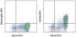
- Staining of 3-day cultured normal human peripheral blood cells with Anti-Human CD14 FITC (Product # 11-0149-42) and Mouse IgG1 K Isotype Control APC (Product # 17-4714-81) (left) or Anti-Human CD169 (Siglec-1) APC (right). Cells in the monocyte gate were used for analysis.

Supportive validation
- Submitted by
- Invitrogen Antibodies (provider)
- Main image
- Experimental details
- NULL
- Submitted by
- Invitrogen Antibodies (provider)
- Main image
- Experimental details
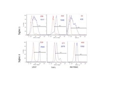
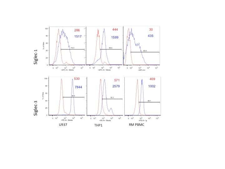
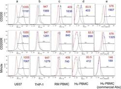
- Fig 2 Representative flow cytometric profiles of CD200, CD200R, Mincle, Siglec-1 and Siglec-3 expression by the U937 (panel a) and THP-1 (panel b) cell lines and by the gated population of CD14+ cells from PBMCs from a representative normal rhesus macaque (panel c) and for comparison the profile of the gated population of CD14+ cells from the human PBMCs stained with our panel of anti-CD200, CD200R and Mincle antibodies (panel d) and the commercially obtained monoclonal antibodies with specificity for human CD200, CD200R and Mincle (panel e). The profile in red reflects the use of anti-mouse Ig (control) and the one in blue reflects the profile noted with the appropriate anti-lectin monoclonal antibody (mAb). The numbers in red and blue are the MFI values noted for the anti-mouse Ig control and the anti-lectin mAb, respectively.

- Submitted by
- Invitrogen Antibodies (provider)
- Main image

- Experimental details
- Fig 3 Representative flow cytometric profile of CD200, CD200R, Mincle, Siglec-1 and Siglec-3 expression on the gated population of CD14 + cells isolated from the bone marrow (panel a), spleen (panel b), liver (panel c) and mononuclear cells isolated from rectal biopsy samples (panel d) from a representative healthy normal rhesus macaque. The profile in red reflects the use of anti-mouse Ig (control) and the one in blue reflects the profile noted with the appropriate anti-lectin monoclonal antibody (mAb). The numbers in red and blue are the MFI values noted for the anti-mouse Ig control and the anti-lectin mAb, respectively.

- Submitted by
- Invitrogen Antibodies (provider)
- Main image

- Experimental details
- Fig. 1 Expression levels of phagocytic receptors on human alveolar macrophages. a FACS gating strategy to delineate alveolar macrophages. Cell suspension was collected by perfusing and lavaging human lung tissues with saline. Alveolar macrophages were detected as live, FSC hi /SSC hi , single cell-gated CD45 + /CD206 + cells in the cell suspension. b Representative histograms for the expression levels of FcgammaRI, CD11b, macrophage scavenger receptor-1 (MSR-1), CD36 and Siglec-1, on FSC hi /SSC hi /CD45 + /CD206 + alveolar macrophages of control never-smokers (CNS, n = 11), control ex-smokers (CES, n = 9) and COPD ex-smokers (COPD, n = 11). Specific staining, open; isotype control, shaded
