 Explore
 Validate
 Learn
Learn Western blot
 Immunohistochemistry
Antibody data
- Antibody Data
- Antigen structure
- References [6]
- Comments [0]
- Validations
- Immunohistochemistry [10]
Submit
Validation data
Reference
Comment
Report error
- Product number
- NB110-74730 - Provider product page

- Provider
- Novus Biologicals
- Proper citation
- Novus Cat#NB110-74730, RRID:AB_1049390
- Product name
- Rabbit Polyclonal Opsin 1 (Medium Wave) Antibody
- Antibody type
- Polyclonal
- Description
- Unpurified.
- Reactivity
- Mouse, Rat
- Host
- Rabbit
- Isotype
- IgG
- Vial size
- 0.1 ml
- Concentration
- LYOPH
- Storage
- Store at 4C short term. Aliquot and store at -20C long term. Avoid freeze-thaw cycles.
Submitted references Novel molecular mechanisms for Prph2-associated pattern dystrophy.
Prph2 initiates outer segment morphogenesis but maturation requires Prph2/Rom1 oligomerization.
JNK1 Induces Notch1 Expression to Regulate Genes Governing Photoreceptor Production.
A Mixture of U.S. Food and Drug Administration-Approved Monoaminergic Drugs Protects the Retina From Light Damage in Diverse Models of Night Blindness.
A Combination of G Protein-Coupled Receptor Modulators Protects Photoreceptors from Degeneration.
The K153Del PRPH2 mutation differentially impacts photoreceptor structure and function.
Chakraborty D, Strayve DG, Makia MS, Conley SM, Kakahel M, Al-Ubaidi MR, Naash MI
FASEB journal : official publication of the Federation of American Societies for Experimental Biology 2020 Jan;34(1):1211-1230
FASEB journal : official publication of the Federation of American Societies for Experimental Biology 2020 Jan;34(1):1211-1230
Prph2 initiates outer segment morphogenesis but maturation requires Prph2/Rom1 oligomerization.
Conley SM, Stuck MW, Watson JN, Zulliger R, Burnett JL, Naash MI
Human molecular genetics 2019 Feb 1;28(3):459-475
Human molecular genetics 2019 Feb 1;28(3):459-475
JNK1 Induces Notch1 Expression to Regulate Genes Governing Photoreceptor Production.
Pan M, Hu H, Wang R, Zhou Y, Zhang L, Wang C, Wang Q
Cells 2019 Aug 24;8(9)
Cells 2019 Aug 24;8(9)
A Mixture of U.S. Food and Drug Administration-Approved Monoaminergic Drugs Protects the Retina From Light Damage in Diverse Models of Night Blindness.
Leinonen H, Choi EH, Gardella A, Kefalov VJ, Palczewski K
Investigative ophthalmology & visual science 2019 Apr 1;60(5):1442-1453
Investigative ophthalmology & visual science 2019 Apr 1;60(5):1442-1453
A Combination of G Protein-Coupled Receptor Modulators Protects Photoreceptors from Degeneration.
Orban T, Leinonen H, Getter T, Dong Z, Sun W, Gao S, Veenstra A, Heidari-Torkabadi H, Kern TS, Kiser PD, Palczewski K
The Journal of pharmacology and experimental therapeutics 2018 Feb;364(2):207-220
The Journal of pharmacology and experimental therapeutics 2018 Feb;364(2):207-220
The K153Del PRPH2 mutation differentially impacts photoreceptor structure and function.
Chakraborty D, Conley SM, Zulliger R, Naash MI
Human molecular genetics 2016 Aug 15;25(16):3500-3514
Human molecular genetics 2016 Aug 15;25(16):3500-3514
No comments: Submit comment
Supportive validation
- Submitted by
- Novus Biologicals (provider)
- Main image
- Experimental details
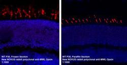
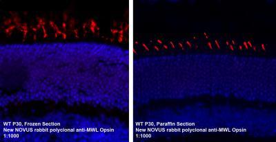
- Immunohistochemistry-Paraffin: Opsin 1 (Medium Wave) Antibody [NB110-74730] - IF analysis of Opsin 1 in paraffin embedded and frozen mouse retina tissues. Image courtesy of product review submitted by Linda Vuong.
- Submitted by
- Novus Biologicals (provider)
- Main image
- Experimental details
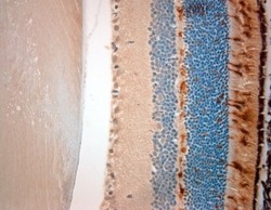
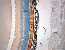
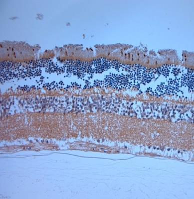
- Immunohistochemistry-Paraffin: Opsin 1 (Medium Wave) Antibody [NB110-74730] - IHC-P on paraffin sections of rat eye. The animal was perfused using Autoperfuser at a pressure of 110 mmHg with 300 ml 4% FA and further post fixed overnight before being processed for paraffin embedding. HIER: Tris-EDTA, pH 9 for 20 min. Blocking: 0.2% LFDM in TBST filtered thru 0.2 um. Detection was done using using HRP polymer following manufacturers inctructions. Primary antibody: dilution 1: 1000, incubated 30 min at RT using Autostainer. Sections were counterstained with Harris Hematoxylin.

- Submitted by
- Novus Biologicals (provider)
- Main image
- Experimental details
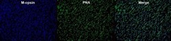
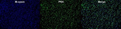
- Immunohistochemistry: Opsin 1 (Medium Wave) Antibody [NB110-74730] - Retinal whole mount of BALB/c mouse stained against anti M-opsin secondary (AF647) antibody and peanut agglutinin (Fluorescein). This image was submitted via customer Review.
- Submitted by
- Novus Biologicals (provider)
- Main image
- Experimental details
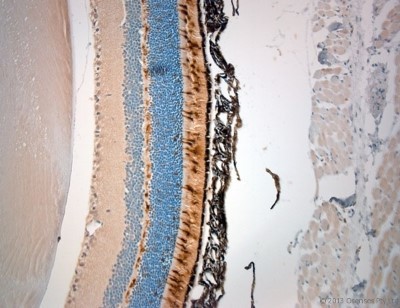
- Immunohistochemistry-Paraffin: Opsin 1 (Medium Wave) Antibody [NB110-74730] - IHC-P on paraffin sections of rat eye.The animal was perfused using Autoperfuser at a pressure of 110 mmHg with 300 ml 4% FA and further post fixed overnight before being processed for paraffin embedding. HIER: Tris-EDTA, pH 9 for 20 min.Blocking: 0.2% LFDM in TBST filtered thru 0.2 um.Detection was done using HRP polymer following manufacturers instructions.Primary antibody: dilution 1: 1000, incubated 30 min at RT using Autostainer.Sections were counterstained with Harris Hematoxylin.
- Submitted by
- Novus Biologicals (provider)
- Main image
- Experimental details
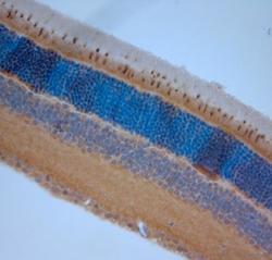
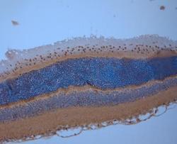
- Immunohistochemistry-Paraffin: Opsin 1 (Medium Wave) Antibody [NB110-74730] - IHC-P on paraffin sections of mouse eye. The animal was perfused at a pressure of 130 mmHg with 300 ml 4% FA before being processed for paraffin embedding. HIER: Tris-EDTA, pH 9 for 20 min. Blocking: 0.2% LFDM in TBST filtered thru 0.2 um. Primary antibody: dilution 1: 500, incubated 30 min at RT. Sections were counterstained with Harris Hematoxylin.
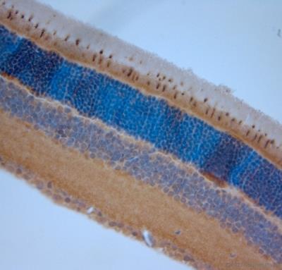
- Submitted by
- Novus Biologicals (provider)
- Main image
- Experimental details
- Immunohistochemistry-Paraffin: Opsin 1 (Medium Wave) Antibody [NB110-74730] - IHC-P on paraffin sections of mouse eye. The animal was perfused at a pressure of 130 mmHg with 300 ml 4% FA before being processed for paraffin embedding. HIER: Tris-EDTA, pH 9 for 20 min. Blocking: 0.2% LFDM in TBST filtered thru 0.2 um. Primary antibody: dilution 1: 500, incubated 30 min at RT. Sections were counterstained with Harris Hematoxylin.

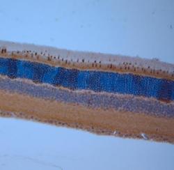
- Submitted by
- Novus Biologicals (provider)
- Main image

- Experimental details
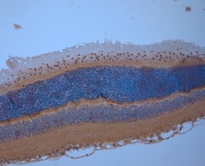
- Immunohistochemistry-Paraffin: Opsin 1 (Medium Wave) Antibody [NB110-74730] - IHC-P on paraffin sections of rat eye. The animal was perfused at a pressure of 130 mmHg with 300 ml 4% FA before being processed for paraffin embedding. HIER: Tris-EDTA, pH 9 for 20 min. Blocking: 0.2% LFDM in TBST filtered thru 0.2 um. Primary antibody: dilution 1: 500, incubated 30 min at RT. Sections were counterstained with Harris Hematoxylin.
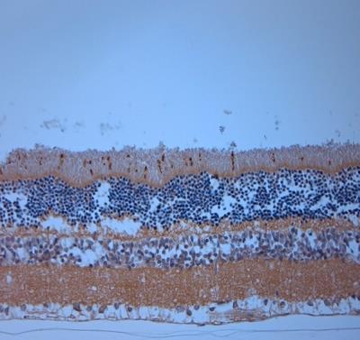
- Submitted by
- Novus Biologicals (provider)
- Main image
- Experimental details
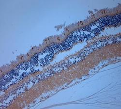
- Immunohistochemistry-Paraffin: Opsin 1 (Medium Wave) Antibody [NB110-74730] - IHC-P on paraffin sections of rat eye. The animal was perfused at a pressure of 130 mmHg with 300 ml 4% FA before being processed for paraffin embedding. HIER: Tris-EDTA, pH 9 for 20 min. Blocking: 0.2% LFDM in TBST filtered thru 0.2 um. Primary antibody: dilution 1: 500, incubated 30 min at RT. Sections were counterstained with Harris Hematoxylin.
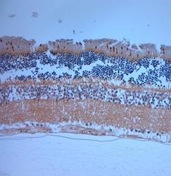
- Submitted by
- Novus Biologicals (provider)
- Main image
- Experimental details
- Immunohistochemistry-Paraffin: Opsin 1 (Medium Wave) Antibody [NB110-74730] - IHC-P on paraffin sections of mouse eye. The animal was perfused at a pressure of 130 mmHg with 300 ml 4% FA before being processed for paraffin embedding. HIER: Tris-EDTA, pH 9 for 20 min. Blocking: 0.2% LFDM in TBST filtered thru 0.2 um. Primary antibody: dilution 1: 500, incubated 30 min at RT. Sections were counterstained with Harris Hematoxylin.
- Submitted by
- Novus Biologicals (provider)
- Main image
- Experimental details
- Immunohistochemistry-Paraffin: Opsin 1 (Medium Wave) Antibody [NB110-74730] - IHC-P on paraffin sections of rat eye. The animal was perfused at a pressure of 130 mmHg with 300 ml 4% FA before being processed for paraffin embedding. HIER: Tris-EDTA, pH 9 for 20 min. Blocking: 0.2% LFDM in TBST filtered thru 0.2 um. Primary antibody: dilution 1: 500, incubated 30 min at RT. Sections were counterstained with Harris Hematoxylin.
