 Explore
 Validate
 Learn
Learn Western blot
Antibody data
- Antibody Data
- Antigen structure
- References [1]
- Comments [0]
- Validations
- Western blot [8]
- Immunocytochemistry [1]
- Immunohistochemistry [1]
- Flow cytometry [1]
- Other assay [1]
Submit
Validation data
Reference
Comment
Report error
- Product number
- PA5-27247 - Provider product page

- Provider
- Invitrogen Antibodies
- Product name
- SQSTM1 Polyclonal Antibody
- Antibody type
- Polyclonal
- Antigen
- Synthetic peptide
- Description
- Recommended positive controls: A549, HepG2, HepG2 (3µM Thapsigargin treatment for 24 hr), Huh7, Huh7 ( 3µM Thapsigargin, 12hr), NIH-3T3, JC, BCL-1, PC-12, Rat2, HeLa.
- Concentration
- 0.39 mg/mL
Submitted references Tau Oligomers and Fibrils Exhibit Differential Patterns of Seeding and Association With RNA Binding Proteins.
Jiang L, Zhao J, Cheng JX, Wolozin B
Frontiers in neurology 2020;11:579434
Frontiers in neurology 2020;11:579434
No comments: Submit comment
Supportive validation
- Submitted by
- Invitrogen Antibodies (provider)
- Main image

- Experimental details
- Western blot analysis of SQSTM1 using 30 µg of A549 lysate. Samples were loaded onto a 10% SDS-PAGE gel and probed with a SQSTM1 polyclonal antibody (Product # PA5-27247) at a dilution of 1:1000.

- Submitted by
- Invitrogen Antibodies (provider)
- Main image

- Experimental details
- Western blot analysis of SQSTM1 using Whole cell extracts (30 µg) prepared from Huh7 cells treated with Thapsigargin for 12 or 24 hours or untreated (-). Samples were loaded onto a 10% SDS-PAGE gel and probed with a SQSTM1 polyclonal antibody (Product # PA5-27247) at a dilution of 1:1000. ACTB was used as an internal control at a dilution of 1:2000, shown at the bottom panel.

- Submitted by
- Invitrogen Antibodies (provider)
- Main image

- Experimental details
- Western Blot analysis of SQSTM1 was performed by separating 30 µg of untreated (–) and treated (+) HepG2 whole cell extracts by 10% SDS-PAGE. Proteins were transferred to a membrane and probed with a SQSTM1 Polyclonal Antibody (Product # PA5-27247) at a dilution of 1:1000.

- Submitted by
- Invitrogen Antibodies (provider)
- Main image

- Experimental details
- Western Blot using SQSTM1 Polyclonal Antibody (Product # PA5-27247). Untreated (–) and treated (+) HepG2 whole cell extracts (30 µg) were separated by 10% SDS-PAGE, and the membrane was blotted with SQSTM1/P62 antibody [N3C1], Internal SQSTM1 Polyclonal Antibody (Product # PA5-27247) diluted at 1:1,000. The HRP-conjugated anti-rabbit IgG antibody was used to detect the primary antibody.

- Submitted by
- Invitrogen Antibodies (provider)
- Main image

- Experimental details
- SQSTM1 antibody [N3C1], Internal detects SQSTM1 protein by western blot analysis. A. 30 µg NIH-3T3 whole cell lysate/extract. B. 30 µg JC whole cell lysate/extract. C. 30 µg BCL-1 whole cell lysate/extract.12% SDS-PAGE. SQSTM1 antibody [N3C1], Internal SQSTM1 Polyclonal Antibody (Product # PA5-27247) dilution: 1:1,000. The HRP-conjugated anti-rabbit IgG antibody was used to detect the primary antibody.

- Submitted by
- Invitrogen Antibodies (provider)
- Main image

- Experimental details
- Western Blot analysis of SQSTM1 was performed by separating 30 µg of untreated (–) and treated (+) Huh-7 whole cell extracts by 10% SDS-PAGE. Proteins were transferred to a membrane and probed with a SQSTM1 Polyclonal Antibody (Product # PA5-27247) at a dilution of 1:1000.

- Submitted by
- Invitrogen Antibodies (provider)
- Main image

- Experimental details
- SQSTM1 antibody [N3C1], Internal detects SQSTM1 protein by western blot analysis. A. 30 µg PC-12 whole cell lysate/extract. B. 30 µg Rat2 whole cell lysate/extract.10% SDS-PAGE. SQSTM1 antibody [N3C1], Internal SQSTM1 Polyclonal Antibody (Product # PA5-27247) dilution: 1:1,000. The HRP-conjugated anti-rabbit IgG antibody was used to detect the primary antibody.

- Submitted by
- Invitrogen Antibodies (provider)
- Main image
- Experimental details
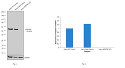
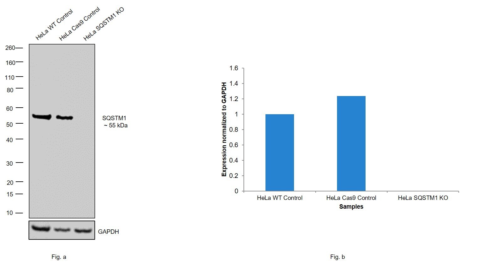
- Knockout of SQSTM1 was achieved by CRISPR-Cas9 genome editing using LentiArray™ Lentiviral sgRNA (Product # A32042, AssayID CRISPR970861_LV) and LentiArray Cas9 Lentivirus (Product # A32064). Western blot analysis of SQSTM1 was performed by loading 30 µg of HeLa wild type (Lane 1), HeLa CAS9 (Lane 2) and HeLa SQSTM1 KO (Lane 3) whole cell extracts. The samples were electrophoresed using Novex® NuPAGE® 4-12% Bis-Tris Protein Gel (Product # NP0321BOX). Resolved proteins were then transferred onto a nitrocellulose membrane (Product # IB23001) by iBlot® 2 Dry Blotting System (Product # IB21001). The blot was probed with Anti-SQSTM1 Polyclonal Antibody (Product # PA5-27247) using 1:1,000 dilution and Goat anti-Rabbit IgG (H+L), Superclonal™ Recombinant Secondary Antibody, HRP (Product # A27036, 1:4,000 dilution) using the iBright FL 1000 (Product # A32752). Chemiluminescent detection was performed using SuperSignal™ West Dura Extended Duration Substrate (Product # 34076). Loss of signal upon CRISPR mediated knockout (KO) using the LentiArray™ CRISPR product line confirms that antibody is specific to SQSTM1.
Supportive validation
- Submitted by
- Invitrogen Antibodies (provider)
- Main image

- Experimental details
- SQSTM1 antibody [N3C1], Internal detects SQSTM1 protein at autophagosome by immunofluorescent analysis. Samples: HeLa cells mock (left) and treated with 50μM Chloroquine for 24 hr (right) were fixed in 4% paraformaldehyde at RT for 15 min. Green: SQSTM1 protein stained by SQSTM1 antibody [N3C1], Internal (Product # PA5-27247) diluted at 1:1,000. Red: Phalloidin, a F-actin marker.

Supportive validation
- Submitted by
- Invitrogen Antibodies (provider)
- Main image

- Experimental details
- SQSTM1/P62 antibody [N3C1], Internal detects SQSTM1/P62 protein at cytoplasm by immunohistochemical analysis. Sample: Paraffin-embedded human lung cancer. SQSTM1/P62 stained by SQSTM1/P62 antibody [N3C1], Internal (Product # PA5-27247) diluted at 1:500. Antigen Retrieval: Citrate buffer, pH 6.0, 15 min.

Supportive validation
- Submitted by
- Invitrogen Antibodies (provider)
- Main image

- Experimental details
- SQSTM1 antibody [N3C1], Internal (Product # PA5-27247) detects SQSTM1 protein by flow cytometry analysis. Sample: HeLa cell fixed in 4% paraformaldehyde at 4°C for 5 min. Brown: Unlabelled sample was also used as a control. Blue: SQSTM1 antibody [N3C1], Internal] (Product # PA5-27247) dilution: 1:100. Acquisition of >20,000 events were collected using Argon ion laser (488nm) and 525/30 bandpass filter.

Supportive validation
- Submitted by
- Invitrogen Antibodies (provider)
- Main image

- Experimental details
- Figure 5 Co-localization of large tau inclusions with p62. (A) Immunoblotting detecting p62 expression in neurons after oTau or fTau treatment. Each lane represents an independent biological replicate. The immunoblotting was performed with cell lysate from hippocampal cultures overexpressing human 4R0N WT tau and treated with vehicle, oTau, or fTau. The cells were harvested at 24 and 96 h after treatment. The p62 antibody was used to identify the expression level of p62 protein. (B) Quantification of p62 immunoblot in cell lysate of hippocampal cultures overexpressing human 4R0N WT tau, treated with vehicle control, oTau, or fTau on day 14, and harvested at 24 or 96 h after treatment. Data are shown as mean +- SEM, N = 4, data analysis was done by two-way ANOVA, multiple comparison test by Fisher's LSD, * p < 0.05, ** p < 0.01 in comparison with vehicle control. (C) Representative images showing the co-localization of phosphorylated tau inclusions CP13 (red) with p62 granules (green) at 96 h after oTau, fTau, or vehicle treatment in hippocampal neurons overexpressing human 4R0N tau. Co-labeling marker is DAPI (blue) to show the cell nucleus. Scale bar 20 mum. (D) Quantification of p62 fluorescence intensity in (C) , which is the immunofluorescence labeling of hippocampal cultures overexpressing human 4R0N WT tau, treated with vehicle control, oTau, or fTau on day 14, and harvested at 96 h after treatment. Data are shown as mean +- SEM, N = 10, data analysis was done by one-wa
