 Explore
 Validate
 Learn
Learn Western blot
 ELISA
ELISAAntibody data
- Antibody Data
- Antigen structure
- References [0]
- Comments [0]
- Validations
- Western blot [1]
- Immunohistochemistry [1]
Submit
Validation data
Reference
Comment
Report error
- Product number
- MA5-14700 - Provider product page

- Provider
- Invitrogen Antibodies
- Product name
- hCG Monoclonal Antibody (3015)
- Antibody type
- Monoclonal
- Antigen
- Other
- Description
- The observed molecular weight of alpha-hCG is approximately 20-22 kD. The molecular weight is higher than the predicted molecular weight due to post translational modification. Product MA514700 is a smaller package size of MIH9805 (formerly sold as a Seradyn product).
- Reactivity
- Human
- Host
- Mouse
- Isotype
- IgG
- Antibody clone number
- 3015
- Vial size
- 100 µg
- Concentration
- 1 mg/mL
- Storage
- Maintain refrigerated at 2-8°C for up to 6 months. For long term storage store at -20°C
No comments: Submit comment
Supportive validation
- Submitted by
- Invitrogen Antibodies (provider)
- Main image

- Experimental details
- Western blot analysis of hCG was performed by loading the indicated amounts of purified hCG protein extracted from human urine, and 15 µL of PageRuler Prestained Protein Ladder (Product # 26619) per well onto a 4-20% Tris-HCl polyacrylamide gel. Proteins were transferred to a PVDF membrane (Product # 88518) using the G2 Fast Blotter (Product # 62288) and blocked with 5% Milk for at least 1 hour at room temperature. hCG was detected at ~22 kD using a anti hCG antibody (Product # MA5-14700) at a concentration of 4 µg/mL in blocking buffer overnight at 4C on a rocking platform, followed by an HRP-conjugated goat anti-mouse IgG (Fc) secondary antibody (Product # 31439) at a dilution of 1:10,000 for at least 1 hour. Chemiluminescent detection was performed using SuperSignal West Dura (Product # 34076).

Supportive validation
- Submitted by
- Invitrogen Antibodies (provider)
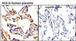
- Main image
- Experimental details
- Immunohistochemistry analysis of hCG was performed on human placenta tissue and is showing positive staining in extracellular spaces. To expose target proteins, antigen retrieval was performed by microwaving tissues for 20 minutes in 10mM sodium citrate buffer (pH 6.0). Tissue slides were probed with a hCG monoclonal antibody (Product # MA5-14700) at a dilution of 1:20, overnight at 4C in a humidified chamber without (right panel) or with hCG antibody (left panel). Tissues were washed, and incubated with secondary antibody (conjugated with HRP) for 30 min at room temperature in a humidified chamber. Detection was performed using DAB substrate kit. Tissues were counterstained with hematoxylin and dehydrated to prep for mounting. Images were taken at 20x magnification.
