PA5-16661
antibody from Invitrogen Antibodies
Targeting: SERPINA1
A1A, A1AT, AAT, alpha-1-antitrypsin, alpha1AT, PI, PI1
Antibody data
- Antibody Data
- Antigen structure
- References [2]
- Comments [0]
- Validations
- Western blot [3]
- Immunocytochemistry [1]
- Immunohistochemistry [1]
- Flow cytometry [1]
- Other assay [3]
Submit
Validation data
Reference
Comment
Report error
- Product number
- PA5-16661 - Provider product page

- Provider
- Invitrogen Antibodies
- Product name
- alpha-1 Antitrypsin Polyclonal Antibody
- Antibody type
- Polyclonal
- Antigen
- Purifed from natural sources
- Description
- PA5-16661 targets Alpha-1-Antitrypsin in IHC (P) applications and shows reactivity with Equine, Human, Mink, monkey, and baboon samples. Does not react with Canine, Chicken, Feline, Guinea Pig, Goat, Marsupial, mouse, Ovine, and Porcine samples
Submitted references Human placental proteomics and exon variant studies link AAT/SERPINA1 with spontaneous preterm birth.
Isolation of adult human pluripotent stem cells from mesenchymal cell populations and their application to liver damages.
Tiensuu H, Haapalainen AM, Tissarinen P, Pasanen A, Määttä TA, Huusko JM, Ohlmeier S, Bergmann U, Ojaniemi M, Muglia LJ, Hallman M, Rämet M
BMC medicine 2022 Apr 28;20(1):141
BMC medicine 2022 Apr 28;20(1):141
Isolation of adult human pluripotent stem cells from mesenchymal cell populations and their application to liver damages.
Wakao S, Kitada M, Kuroda Y, Dezawa M
Methods in molecular biology (Clifton, N.J.) 2012;826:89-102
Methods in molecular biology (Clifton, N.J.) 2012;826:89-102
No comments: Submit comment
Supportive validation
- Submitted by
- Invitrogen Antibodies (provider)
- Main image
- Experimental details
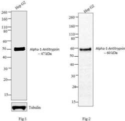
- Western blot analysis was performed on membrane enriched extracts (30 µg) of (Fig 1) Hep G2 (Lane 1). Likewise, Western blot analysis was performed on (Fig 2) condition media of Hep G2 (Lane 1).The blots were probed with Anti-Alpha1-Antitrypsin Rabbit Polyclonal Antibody (Product # PA5-16661, 1:250 dilution) and detected by chemiluminescence using Goat anti-Rabbit IgG (H+L) Superclonal™ Secondary Antibody, HRP conjugate (Product # A27036, 0.4 µg/mL, 1:2500 dilution) A ~47 kDa band corresponding Alpha1-Antitrypsin was observed in Hep G2 (Fig 1) and a higher molecular weight of 60 kDa was observed in Hep G2 conditioned media (Fig 2) which is due to glycosylation of the protein. Known quantity of protein samples were electrophoresed using Novex® NuPAGE®4-12 % Bis-Tris gel (Product # NP0321BOX), XCell SureLock™ Electrophoresis System (Product # EI0002) and Novex® Sharp Pre-Stained Protein Standard (Product # LC5800). Resolved proteins were then transferred onto a nitrocellulose membrane by iBlot® 2 Dry Blotting System (Product # IB21001). The membrane was probed with the relevant primary and secondary Antibody using iBind™ Flex Western Starter Kit (Product # SLF2000S). Chemiluminescent detection was performed using Pierce™ ECL Western Blotting Substrate (Product # 32106).
- Submitted by
- Invitrogen Antibodies (provider)
- Main image
- Experimental details
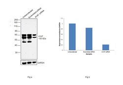
- Knockdown of alpha-1 Antitrypsin was achieved by transfecting Hep G2 with alpha-1 Antitrypsin specific siRNAs (Silencer® select Product # S10459, S10458). Western blot analysis (Fig. a) was performed using Membrane enriched extracts from the alpha-1 Antitrypsin knockdown cells (lane 3), non-targeting scrambled siRNA transfected cells (lane 2) and untransfected cells (lane 1). The blot was probed with alpha-1 Antitrypsin Polyclonal Antibody (Product # PA5-16661, 1:250 dilution) and Goat anti-Rabbit IgG (H+L) Superclonal™ Recombinant Secondary Antibody, HRP (Product # A27036, 1:4000 dilution). Densitometric analysis of this western blot is shown in histogram (Fig. b). Decrease in signal upon siRNA mediated knock down confirms that antibody is specific to alpha-1 Antitrypsin.
- Submitted by
- Invitrogen Antibodies (provider)
- Main image
- Experimental details
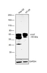
- Western blot was performed using Anti-alpha-1 Antitrypsin Polyclonal Antibody(Product # PA5-16661) and a 55kDa band corresponding to alpha-1 Antitrypsin was observed across cell lines tested. Membrane enriched extracts (30 µg lysate) of Hep G2 (Lane 1) and HT-29 (Lane 2) were electrophoresed using NuPAGE™ 10% Bis-Tris Protein Gel (Product # NP0301BOX). Resolved proteins were then transferred onto a Nitrocellulose membrane (Product # IB23001) by iBlot® 2 Dry Blotting System (Product # IB21001). The blot was probed with the primary antibody (1:250 dilution) and detected by chemiluminescence with Goat anti-Rabbit IgG (H+L) Superclonal™ Recombinant Secondary Antibody, HRP (Product # A27036,1:4000 dilution) using the iBright FL 1000 (Product # A32752). Chemiluminescent detection was performed using Novex® ECL Chemiluminescent Substrate Reagent Kit (Product # WP20005).
Supportive validation
- Submitted by
- Invitrogen Antibodies (provider)
- Main image
- Experimental details
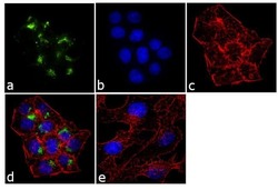
- Immunofluorescent analysis of Alpha-1-Antitrypsin was performed using 70% confluent log phase Hep G2 cells. The cells were fixed with 4% paraformaldehyde for 10 minutes, permeabilized with 0.1% Triton™ X-100 for 10 minutes, and blocked with 1% BSA for 1 hour at room temperature. The cells were labeled with Alpha-1-Antitrypsin Rabbit Polyclonal Antibody (Product # PA5-16661) at 1:250 dilution in 0.1% BSA and incubated for 3 hours at room temperature and then labeled with Goat anti-Rabbit IgG (H+L) Superclonal™ Secondary Antibody, Alexa Fluor® 488 conjugate (Product # A27034) a dilution of 1:2000 for 45 minutes at room temperature (Panel a: green). Nuclei (Panel b: blue) were stained with SlowFade® Gold Antifade Mountant with DAPI (Product # S36938). F-actin (Panel c: red) was stained with Alexa Fluor® 555 Rhodamine Phalloidin (Product # R415, 1:300). Panel d represents the merged image showing cytoplasmic localization. Panel e shows the no primary antibody control. The images were captured at 60X magnification.
Supportive validation
- Submitted by
- Invitrogen Antibodies (provider)
- Main image
- Experimental details
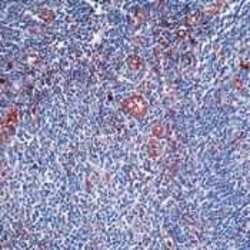
- Formalin-fixed, paraffin-embedded human tonsil stained with Alpha-1ntitrypsin antibody using peroxidase-conjugate and AEC chromogen. Note cytoplasmic staining of endothelial cells.
Supportive validation
- Submitted by
- Invitrogen Antibodies (provider)
- Main image
- Experimental details
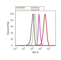
- Flow cytometry analysis of Alpha-1-Antitrypsin was done on Hep G2 cells. Cells were fixed with 70% ethanol for 10 minutes, permeabilized with 0.25% Triton™ X-100 for 20 minutes, and blocked with 5% BSA for 30 minutes at room temperature. Cells were labeled with Alpha-1-Antitrypsin Rabbit Polyclonal Antibody (PA5-16661, red histogram) or with rabbit isotype control (pink histogram) at 3-5 ug/million cells in 2.5% BSA. After incubation at room temperature for 2 hours, the cells were labeled with Alexa Fluor® 488 Goat Anti-Rabbit Secondary Antibody (A11008) at a dilution of 1:400 for 30 minutes at room temperature. The representative 10, 000 cells were acquired and analyzed for each sample using an Attune® Acoustic Focusing Cytometer. The purple histogram represents unstained control cells and the green histogram represents no-primary-antibody control.
Supportive validation
- Submitted by
- Invitrogen Antibodies (provider)
- Main image

- Experimental details
- Placental localization of AAT in spontaneous preterm (SPTB) and term (STB) births. Samples from SPTB and STB placentas were immunostained with anti-human AAT antibody. Samples were from the basal plate (maternal side) and from the decidua of the placenta. Immunostaining is indicated by arrowheads in villous trophoblasts (cytotrophoblasts and syncytiotrophoblasts) and arrows in decidual trophoblasts. Original magnification x 20 in all figures. Control is isotype control for immunostaining. Scale bar: 100 mum
- Submitted by
- Invitrogen Antibodies (provider)
- Main image
- Experimental details
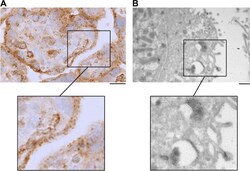
- Stained cytoplasmic AAT granules in syncytiotrophoblasts of spontaneous term birth placenta. In both A and B , samples from the basal plate (maternal side) of the placenta immunostained with anti-human AAT antibody. A Immunohistochemistry image of AAT staining in syncytiotrophoblasts. Scale bar: 20 mum. B Immunoelectron microscopy image of AAT staining in syncytiotrophoblasts. Scale bar: 0.5 mum
- Submitted by
- Invitrogen Antibodies (provider)
- Main image
- Experimental details
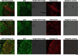
- Confocal microscopy images of human placenta expressing endogenous AAT, fibronectin, vitronectin, and collagen IV. Colocalization fluorescence image of AAT (green) and fibronectin (red) (top row), AAT (green) and vitronectin (red) (middle row), and AAT (green) and collagen IV (red) (bottom row). Middle columns are separate green channel, red channel, and brightfield images, which are shown merged in left column. Panels in right column show negative controls treated the same way as samples but with primary antibody omitted. Objective lens: HC PL APO CS2 x 20/0.75 DRY. Scale bar: 10 mum
 Explore
 Validate
 Learn
Learn Western blot