 Explore
 Validate
 Learn
Learn Immunocytochemistry
 Immunohistochemistry
Antibody data
- Antibody Data
- Antigen structure
- References [0]
- Comments [0]
- Validations
- Immunocytochemistry [1]
- Immunohistochemistry [5]
Submit
Validation data
Reference
Comment
Report error
- Product number
- HPA001223 - Provider product page

- Provider
- Atlas Antibodies
- Proper citation
- Atlas Antibodies Cat#HPA001223, RRID:AB_1846112
- Product name
- Anti-CBS
- Antibody type
- Polyclonal
- Reactivity
- Human
- Host
- Rabbit
- Conjugate
- Unconjugated
- Antigen sequence
GGSVKDRISLRMIEDAERDGTLKPGDTIIEPTSGN
TGIGLALAAAVRGYRCIIVMPEKMSSEKVDVLRAL
GAEIVRTPTNARFDSPESHVGVAWRLKNEIPNSHI
LDQYRNASNPLAHYDTTADEILQQCDGKLDMLVAS
VGTGGTITGIGAPHR- Isotype
- IgG
- Vial size
- 100 µl
- Storage
- Store at +4°C for short term storage. Long time storage is recommended at -20°C.
No comments: Submit comment
Supportive validation
- Submitted by
- Atlas Antibodies (provider)
- Main image

- Experimental details
- Immunofluorescent staining of human cell line U-251 MG shows localization to nucleoli & vesicles.
- Sample type
- HUMAN

Enhanced validation
Supportive validation
- Submitted by
- Atlas Antibodies (provider)
- Enhanced method
- Orthogonal validation
- Main image

- Experimental details
- Immunohistochemistry analysis in human liver and skeletal muscle tissues using HPA001223 antibody. Corresponding CBS RNA-seq data are presented for the same tissues.
- Sample type
- HUMAN

Supportive validation
- Submitted by
- Atlas Antibodies (provider)
- Main image

- Experimental details
- Immunohistochemical staining of human liver shows moderate cytoplasmic positivity in hepatocytes.
- Sample type
- HUMAN

- Submitted by
- Atlas Antibodies (provider)
- Main image
- Experimental details
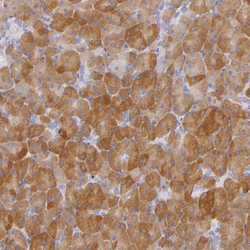
- Immunohistochemical staining of human pancreas shows moderate cytoplasmic positivity in exocrine glandular cells.
- Sample type
- HUMAN

- Submitted by
- Atlas Antibodies (provider)
- Main image

- Experimental details
- Immunohistochemical staining of human cerebellum shows moderate cytoplasmic positivity in a subset of cells in granular layer, as well as in neuropil of molecular layer.
- Sample type
- HUMAN

- Submitted by
- Atlas Antibodies (provider)
- Main image

- Experimental details
- Immunohistochemical staining of human skeletal muscle shows no positivity in striated muscle fibers as expected.
- Sample type
- HUMAN
