Explore
 Validate
 Learn
Learn Western blot
Antibody data
- Antibody Data
- Antigen structure
- References [1]
- Comments [0]
- Validations
- Western blot [3]
- Immunohistochemistry [1]
Submit
Validation data
Reference
Comment
Report error
- Product number
- PA5-81948 - Provider product page

- Provider
- Invitrogen Antibodies
- Product name
- ECE1 Polyclonal Antibody
- Antibody type
- Polyclonal
- Antigen
- Recombinant full-length protein
- Reactivity
- Human
- Host
- Rabbit
- Isotype
- IgG
- Vial size
- 100 µL
- Concentration
- 0.05 mg/mL
- Storage
- Store at 4°C short term. For long term storage, store at -20°C, avoiding freeze/thaw cycles.
Submitted references Induction of reactive oxygen species by mechanical stretch drives endothelin production in neonatal pig renal epithelial cells.
Kumar R, Soni H, Afolabi JM, Kanthakumar P, Mankuzhy PD, Iwhiwhu SA, Adebiyi A
Redox biology 2022 Jul 4;55:102394
Redox biology 2022 Jul 4;55:102394
No comments: Submit comment
Supportive validation
- Submitted by
- Invitrogen Antibodies (provider)
- Main image
- Experimental details
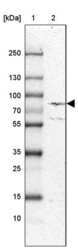
- Western blot analysis of ECE1 by a ECE1 polyclonal antibody (Product # PA5-81948). Lane 1: Marker [kDa] 250, 130, 100, 70, 55, 35, 25, 15, 10 Lane 2: Human cell line U-87 MG.
- Submitted by
- Invitrogen Antibodies (provider)
- Main image

- Experimental details
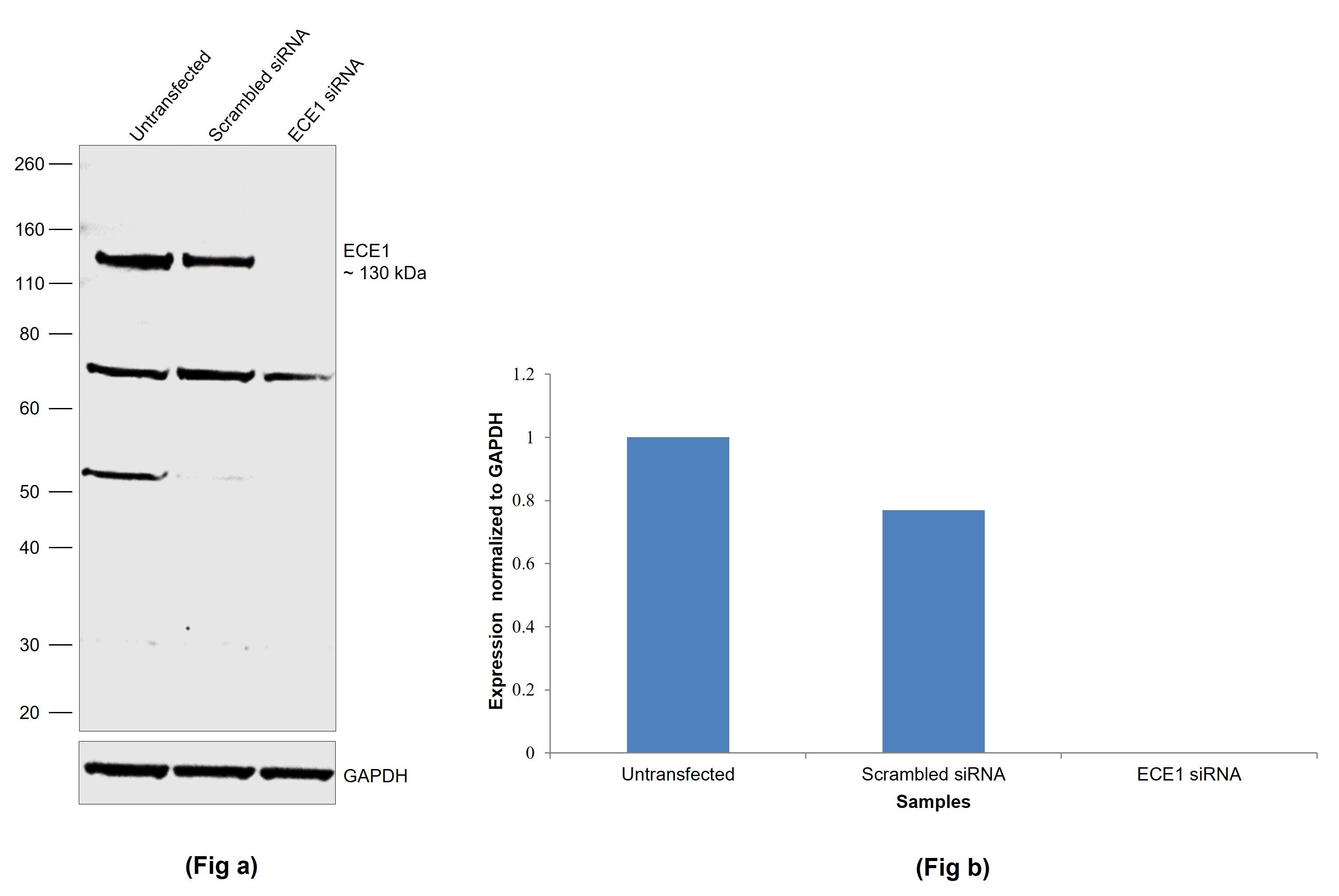
- Knockdown of ECE1 was achieved by transfecting HeLa with ECE1 specific siRNAs (Silencer® select Product # s644, s646). Western blot analysis (Fig. a) was performed using whole cell extracts from the ECE1 knockdown cells (lane 3), non-targeting scrambled siRNA transfected cells (lane 2) and untransfected cells (lane 1). The blot was probed with ECE1 Polyclonal Antibody (Product # PA5-81948, 0.1 µg/mL) and Goat anti-Rabbit IgG (H+L) Superclonal™ Recombinant Secondary Antibody, HRP (Product # A27036, 1:20,000) and detected by chemiluminescence using the iBright™ FL1500 Imaging System (Product # A44115). Densitometric analysis of this western blot is shown in histogram (Fig. b). Decrease in signal upon siRNA mediated knock down confirms that antibody is specific to ECE1.
- Submitted by
- Invitrogen Antibodies (provider)
- Main image
- Experimental details
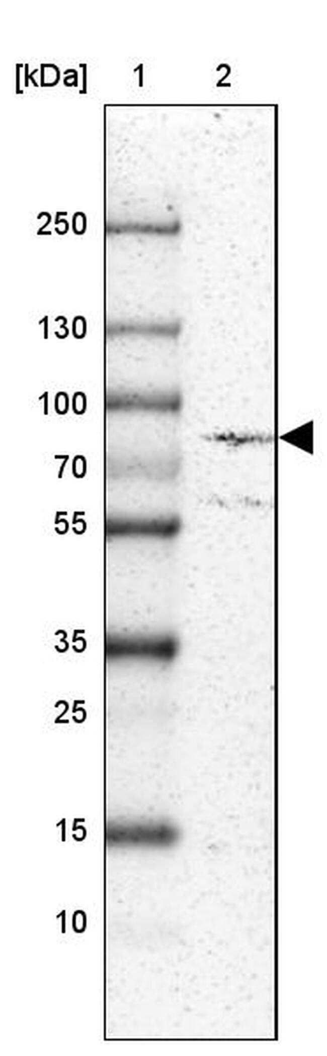
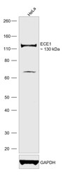
- Western blot was performed using ECE1 Polyclonal Antibody (Product # PA5-81948) and a 130 kDa band corresponding to ECE1 was observed. Whole cell extract (50 µg lysate) of HeLa was electrophoresed using NuPAGE™ 4-12% Bis-Tris Protein Gel (Product # NP0322BOX), 12 well. Resolved proteins were then transferred onto a nitrocellulose membrane (Product # IB23001) by iBlot® 2 Dry Blotting System (Product # IB21001). The blot was probed with the primary antibody (0.1 µg/mL) and detected by chemiluminescence with Goat anti-Rabbit IgG (H+L) Superclonal™ Recombinant Secondary Antibody, HRP (Product # A27036, 1:20,000) using the iBright™ FL1500 Imaging System (Product # A44115). Chemiluminescent detection was performed using SuperSignal™ West Pico PLUS Chemiluminescent Substrate (Product # 34580).

Supportive validation
- Submitted by
- Invitrogen Antibodies (provider)
- Main image

- Experimental details
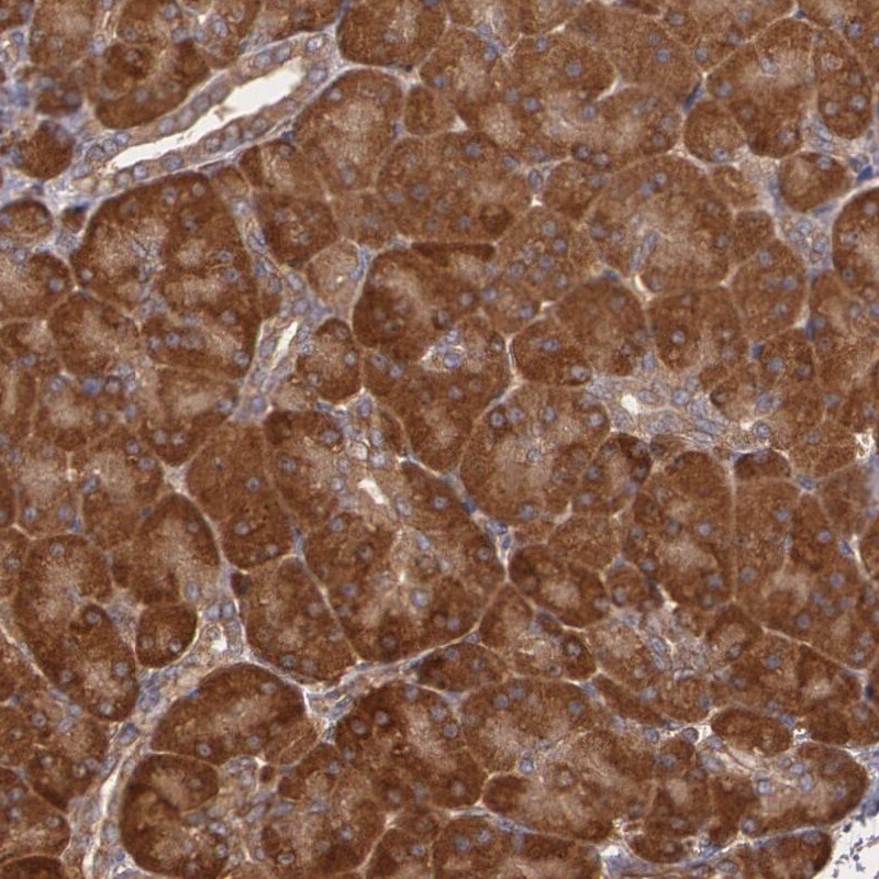
- Immunohistochemical analysis of ECE1 in human pancreas using a ECE1 polyclonal antibody (Product # PA5-81948). The analysis shows strong cytoplasmic positivity in exocrine glandular cells.