 Explore
 Validate
 Learn
Learn Western blot
 Immunoprecipitation
Antibody data
- Antibody Data
- Antigen structure
- References [6]
- Comments [0]
- Validations
- Western blot [7]
- Immunocytochemistry [2]
- Immunohistochemistry [5]
- Other assay [4]
Submit
Validation data
Reference
Comment
Report error
- Product number
- PA5-85197 - Provider product page

- Provider
- Invitrogen Antibodies
- Product name
- MMP2 Polyclonal Antibody
- Antibody type
- Polyclonal
- Antigen
- Recombinant full-length protein
- Reactivity
- Human
- Host
- Rabbit
- Isotype
- IgG
- Vial size
- 100 µL
- Concentration
- 0.44 mg/mL
- Storage
- Store at 4°C short term. For long term storage, store at -20°C, avoiding freeze/thaw cycles.
Submitted references TRIM66 Promotes Malignant Progression of Non-Small-Cell Lung Cancer Cells via Targeting MMP9.
Poria Acid, Triterpenoids Extracted from Poria cocos, Inhibits the Invasion and Metastasis of Gastric Cancer Cells.
Betulonic Acid, as One of the Active Components of the Celastrus orbiculatus Extract, Inhibits the Invasion and Metastasis of Gastric Cancer Cells by Mediating Cytoskeleton Rearrangement In Vitro.
Cardiac Protective Effect of Kirenol against Doxorubicin-Induced Cardiac Hypertrophy in H9c2 Cells through Nrf2 Signaling via PI3K/AKT Pathways.
Chondroitin polymerizing factor promotes breast carcinoma cell proliferation, invasion and migration and affects expression of epithelial-mesenchymal transition-related markers.
Coronin 1C inhibits melanoma metastasis through regulation of MT1-MMP-containing extracellular vesicle secretion.
Xu Y, Yang Q, Fang Z, Tan X, Zhang M, Chen W
Computational and mathematical methods in medicine 2022;2022:6058720
Computational and mathematical methods in medicine 2022;2022:6058720
Poria Acid, Triterpenoids Extracted from Poria cocos, Inhibits the Invasion and Metastasis of Gastric Cancer Cells.
Wang H, Luo Y, Chu Z, Ni T, Ou S, Dai X, Zhang X, Liu Y
Molecules (Basel, Switzerland) 2022 Jun 6;27(11)
Molecules (Basel, Switzerland) 2022 Jun 6;27(11)
Betulonic Acid, as One of the Active Components of the Celastrus orbiculatus Extract, Inhibits the Invasion and Metastasis of Gastric Cancer Cells by Mediating Cytoskeleton Rearrangement In Vitro.
Chu Z, Luo Y, Ni T, Zhu M, Feng X, Liu Y, Wang H
Molecules (Basel, Switzerland) 2022 Feb 2;27(3)
Molecules (Basel, Switzerland) 2022 Feb 2;27(3)
Cardiac Protective Effect of Kirenol against Doxorubicin-Induced Cardiac Hypertrophy in H9c2 Cells through Nrf2 Signaling via PI3K/AKT Pathways.
Alzahrani AM, Rajendran P, Veeraraghavan VP, Hanieh H
International journal of molecular sciences 2021 Mar 23;22(6)
International journal of molecular sciences 2021 Mar 23;22(6)
Chondroitin polymerizing factor promotes breast carcinoma cell proliferation, invasion and migration and affects expression of epithelial-mesenchymal transition-related markers.
Li Y, Gong H, Feng L, Mao D, Xiao Y, Wang Y, Huang L
FEBS open bio 2021 Feb;11(2):423-434
FEBS open bio 2021 Feb;11(2):423-434
Coronin 1C inhibits melanoma metastasis through regulation of MT1-MMP-containing extracellular vesicle secretion.
Tagliatela AC, Hempstead SC, Hibshman PS, Hockenberry MA, Brighton HE, Pecot CV, Bear JE
Scientific reports 2020 Jul 20;10(1):11958
Scientific reports 2020 Jul 20;10(1):11958
No comments: Submit comment
Supportive validation
- Submitted by
- Invitrogen Antibodies (provider)
- Main image

- Experimental details
- Western blot analysis of MMP2 in various whole cell extracts using MMP2 polyclonal antibody (Product # PA5-85197) using 30 µg of sample at a dilution of 1:500. Sample was then incubated with HRP-conjugated anti-rabbit IgG secondary antibody. Prior to incubation with primary antibody, the sample was separated on 7.5% SDS-PAGE.

- Submitted by
- Invitrogen Antibodies (provider)
- Main image

- Experimental details
- Western blot analysis of MMP2 in various whole cell extracts using MMP2 polyclonal antibody (Product # PA5-85197) using 30 µg of sample at a dilution of 1:3000. Sample was then incubated with HRP-conjugated anti-rabbit IgG secondary antibody. Prior to incubation with primary antibody, the sample was separated on 7.5% SDS-PAGE.

- Submitted by
- Invitrogen Antibodies (provider)
- Main image

- Experimental details
- Western blot analysis of MMP2 in U87-MG whole cell extract using MMP2 polyclonal antibody (Product # PA5-85197) using 30 µg of sample at a dilution of 1:500. Sample was then incubated with HRP-conjugated anti-rabbit IgG secondary antibody. Prior to incubation with primary antibody, the sample was separated on 7.5% SDS-PAGE.

- Submitted by
- Invitrogen Antibodies (provider)
- Main image

- Experimental details
- Western blot analysis of MMP2 in A) mouse brain using MMP2 polyclonal antibody (Product # PA5-85197) using 50 µg of sample at a dilution of 1:1000. Sample was then incubated with HRP-conjugated anti-rabbit IgG secondary antibody. Prior to incubation with primary antibody, the sample was separated on 7.5% SDS-PAGE.

- Submitted by
- Invitrogen Antibodies (provider)
- Main image

- Experimental details
- Western Blot analysis of MMP2 was performed by separating 30 µg of Whole cell extracts by 7.5% SDS-PAGE. Proteins were transferred to a membrane and probed with a MMP2 Polyclonal Antibody (Product # PA5-85197) at a dilution of 1:1000. The HRP-conjugated anti-rabbit IgG antibody was used to detect the primary antibody.

- Submitted by
- Invitrogen Antibodies (provider)
- Main image

- Experimental details
- Western Blot using MMP2 Polyclonal Antibody (Product # PA5-85197). Whole cell extract (30 µg) was separated by 7.5% SDS-PAGE, and the membrane was blotted with MMP2 Polyclonal Antibody (Product # PA5-85197) diluted at 1:1,000. The HRP-conjugated anti-rabbit IgG antibody was used to detect the primary antibody.

- Submitted by
- Invitrogen Antibodies (provider)
- Main image

- Experimental details
- Western Blot using MMP2 Polyclonal Antibody (Product # PA5-85197). U87-MG whole cell extract and conditioned medium (30 µg) were separated by 7.5% SDS-PAGE, and the membrane was blotted with MMP2 Polyclonal Antibody (Product # PA5-85197) diluted at 1:4,000. The HRP-conjugated anti-rabbit IgG antibody was used to detect the primary antibody.

Supportive validation
- Submitted by
- Invitrogen Antibodies (provider)
- Main image
- Experimental details
- Immunocytochemistry analysis of MMP2 in HeLa cells using MMP2 polyclonal antibody (Product # PA5-85197) at a dilution of 1:200.

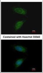
- Submitted by
- Invitrogen Antibodies (provider)
- Main image

- Experimental details
- MMP2 Polyclonal Antibody detects MMP2 protein at cytoplasm by immunofluorescent analysis. Sample: U87-MG cells were fixed in 4% paraformaldehyde at RT for 15 min. Green: MMP2 stained by MMP2 Polyclonal Antibody (Product # PA5-85197) diluted at 1:500. Red: alpha Tubulin, a cytoskeleton marker, stained by alpha Tubulin Polyclonal Antibody [GT114] (Product # MA5-31466) diluted at 1:1,000. Blue: Fluoroshield with DAPI .

Supportive validation
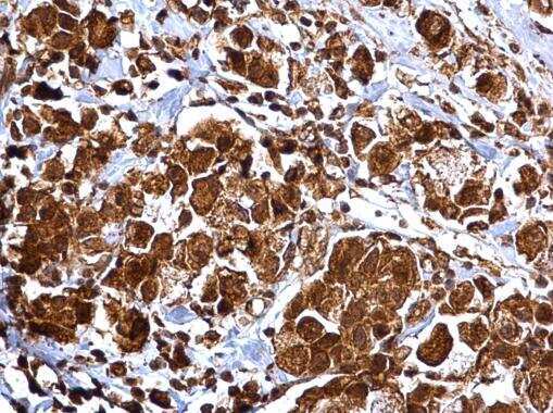
- Submitted by
- Invitrogen Antibodies (provider)
- Main image

- Experimental details
- MMP2 Polyclonal Antibody detects MMP2 protein at cytosol and nucleus on human ovarian carcinoma by immunohistochemical analysis. Sample: Paraffin-embedded human ovarian carcinoma. MMP2 Polyclonal Antibody (Product # PA5-85197) dilution: 1:500. Antigen Retrieval: EDTA based buffer, pH 8.0, 15 min.
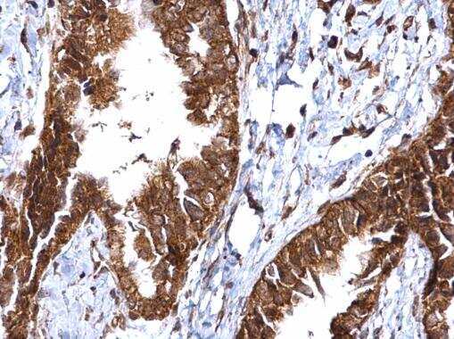
- Submitted by
- Invitrogen Antibodies (provider)
- Main image

- Experimental details
- MMP2 Polyclonal Antibody detects MMP2 protein at extracellular matrix in human lung cancer by immunohistochemical analysis. Sample: Paraffin-embedded human lung cancer. MMP2 Polyclonal Antibody (Product # PA5-85197) diluted at 1:500. Antigen Retrieval: Citrate buffer, pH 6.0, 15 min.

- Submitted by
- Invitrogen Antibodies (provider)
- Main image

- Experimental details
- MMP2 Polyclonal Antibody detects MMP2 protein at cytosol on human endometrial carcinoma by immunohistochemical analysis. Sample: Paraffin-embedded human endometrial carcinoma. MMP2 Polyclonal Antibody (Product # PA5-85197) dilution: 1:500. Antigen Retrieval: EDTA based buffer, pH 8.0, 15 min.
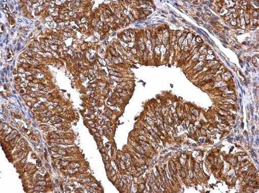
- Submitted by
- Invitrogen Antibodies (provider)
- Main image

- Experimental details
- MMP2 Polyclonal Antibody detects MMP2 protein at extracellular matrix in human colon cancer by immunohistochemical analysis. Sample: Paraffin-embedded human colon cancer. MMP2 Polyclonal Antibody (Product # PA5-85197) diluted at 1:500. Antigen Retrieval: Citrate buffer, pH 6.0, 15 min.

- Submitted by
- Invitrogen Antibodies (provider)
- Main image

- Experimental details
- MMP2 Polyclonal Antibody detects MMP2 protein at cytosol and nucleus on human breast carcinoma by immunohistochemical analysis. Sample: Paraffin-embedded human breast carcinoma. MMP2 Polyclonal Antibody (Product # PA5-85197) dilution: 1:500. Antigen Retrieval: EDTA based buffer, pH 8.0, 15 min.
Supportive validation
- Submitted by
- Invitrogen Antibodies (provider)
- Main image

- Experimental details
- Immunoprecipitation analysis of MMP2 in U87-MG whole cell extracts with MMP2 polyclonal antibody (Product # PA5-85197) using 5 µg of sample, followed by anti-Rabbit IgG secondary antibody.

- Submitted by
- Invitrogen Antibodies (provider)
- Main image

- Experimental details
- Figure 2 Effects of KRL on DOX-induced hypertrophy. H9c2 cells were treated for 24 h with 15 uM of KRL, 2 h before DOX (0.25 umol) treatment. ( A ) Representative Western blots showing changes in the protein levels of ANP and BNP. ( B ) KRL is illustrated to downregulate MMP9 and MMP2. Western blot analysis was performed to determine the total protein MMP9 and MMp2 levels in the total extract by including beta-actin as an internal loading control. ( C ) H9c2 cells were treated for 24 h with 10 and 15 uM of KRL, 2 h before the DOX (0.25 umol) treatment. The cells then underwent actin filament staining to observe the changes in the surface area of the cardiomyocytes. Scale bar indicated 100u m at 20x magnification. Data are represented as the mean +- SD of triplicate values ( n = 3) and * p < 0.05 represents significant variations compared with the control. # p < 0.05 represents significant variations as compared to DOX alone and KRL with DOX treatment groups.

- Submitted by
- Invitrogen Antibodies (provider)
- Main image

- Experimental details
- 5 Fig. CHPF is associated with EMT relative markers and MMP2. (A-D) The GEPIA database was used to analyze the relationship between CHPF and metastatic markers, including vimentin, Snail, Slug and MMP2. (0 < r < 1, Pearson correlation coefficient). (E, F) Effects of CHPF depletion (E) or overexpression (F) on motion-related markers in MCF7 (E) or MDA-MB-231 (F) cells were determined by western blotting. ** P < 0.01 vs. si-con group or vector group. Data are presented as the mean +- SD for three independent experiments. Statistical analyses were performed using Student's t test.

- Submitted by
- Invitrogen Antibodies (provider)
- Main image
- Experimental details
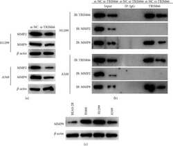
- The bindings between TRIM66 and MMP2 or MMP9. (a) Protein levels of MMP2 and MMP9 in each transfection group. (b) Cells were lysed and immunoprecipitated with antibodies, and immunocomplexes were analyzed by Western blot. (c) MMP9 level in proteins extracted from NSCLC and pulmonary alveolar epithelial cells.
