Explore
 Validate
 Learn
Learn Western blot
 ELISA
ELISA Immunocytochemistry
Antibody data
- Antibody Data
- Antigen structure
- References [26]
- Comments [0]
- Validations
- Western blot [11]
- Immunocytochemistry [1]
- Immunoprecipitation [2]
- Immunohistochemistry [2]
- Flow cytometry [1]
Submit
Validation data
Reference
Comment
Report error
- Product number
- GTX109242 - Provider product page

- Provider
- GeneTex
- Proper citation
- GeneTex Cat#GTX109242, RRID:AB_2036264
- Product name
- Arginase 1 antibody
- Antibody type
- Polyclonal
- Reactivity
- Human, Mouse, Rat
- Host
- Rabbit
Submitted references HYCO-3, a dual CO-releaser/Nrf2 activator, reduces tissue inflammation in mice challenged with lipopolysaccharide.
Lysed Erythrocyte Membranes Promote Vascular Calcification.
Interleukin-1β has atheroprotective effects in advanced atherosclerotic lesions of mice.
A RIPK3-PGE2 Circuit Mediates Myeloid-Derived Suppressor Cell-Potentiated Colorectal Carcinogenesis.
Increased number of arginase 1-positive cells in the stroma of carcinomas compared to precursor lesions and nonneoplastic tissues.
Langerhans, plasmacytoid dendritic and myeloid-derived suppressor cell levels in mycosis fungoides vary according to the stage of the disease.
Galectin-1 Reduces Neuroinflammation via Modulation of Nitric Oxide-Arginase Signaling in HIV-1 Transfected Microglia: a Gold Nanoparticle-Galectin-1 "Nanoplex" a Possible Neurotherapeutic?
Metabolic origins of spatial organization in the tumor microenvironment.
Hepatocyte-secreted extracellular vesicles modify blood metabolome and endothelial function by an arginase-dependent mechanism.
Roles of programmed death protein 1/programmed death-ligand 1 in secondary brain injury after intracerebral hemorrhage in rats: selective modulation of microglia polarization to anti-inflammatory phenotype.
Identification of different macrophage subpopulations with distinct activities in a mouse model of oxygen-induced retinopathy.
IRAK-M alters the polarity of macrophages to facilitate the survival of Mycobacterium tuberculosis.
The prognostic value of the myeloid-mediated immunosuppression marker Arginase-1 in classic Hodgkin lymphoma.
Cyclic AMP is a key regulator of M1 to M2a phenotypic conversion of microglia in the presence of Th2 cytokines.
MicroRNA-200c Promotes Suppressive Potential of Myeloid-Derived Suppressor Cells by Modulating PTEN and FOG2 Expression.
Poly(I:C) and CpG-ODN combined aerosolization to treat lung metastases and counter the immunosuppressive microenvironment.
Tissue matrix arrays for high-throughput screening and systems analysis of cell function.
Fat-laden macrophages modulate lobular inflammation in nonalcoholic steatohepatitis (NASH).
Arterial dysfunction but maintained systemic blood pressure in cavin-1-deficient mice.
The omega-3 fatty acid docosahexaenoic acid favorably modulates the inflammatory pathways and macrophage polarization within aorta of LDLR(-/-) mice.
Macrophage-derived apoESendai suppresses atherosclerosis while causing lipoprotein glomerulopathy in hyperlipidemic mice.
Anti-inflammatory immune skewing is atheroprotective: Apoe−/−FcγRIIb−/− mice develop fibrous carotid plaques.
Role of uterine contraction in regeneration of the murine postpartum endometrium.
Inhibition of endogenous activated protein C attenuates experimental autoimmune encephalomyelitis by inducing myeloid-derived suppressor cells.
Elevated pulmonary arterial pressure and altered expression of Ddah1 and Arg1 in mice lacking cavin-1/PTRF.
Antisense oligonucleotide treatment enhances the recovery of acute lung injury through IL-10-secreting M2-like macrophage-induced expansion of CD4+ regulatory T cells.
Motterlini R, Nikam A, Manin S, Ollivier A, Wilson JL, Djouadi S, Muchova L, Martens T, Rivard M, Foresti R
Redox biology 2019 Jan;20:334-348
Redox biology 2019 Jan;20:334-348
Lysed Erythrocyte Membranes Promote Vascular Calcification.
Tziakas DN, Chalikias G, Pavlaki M, Kareli D, Gogiraju R, Hubert A, Böhm E, Stamoulis P, Drosos I, Kikas P, Mikroulis D, Giatromanolaki A, Georgiadis GS, Konstantinou F, Argyriou C, Münzel T, Konstantinides SV, Schäfer K
Circulation 2019 Apr 23;139(17):2032-2048
Circulation 2019 Apr 23;139(17):2032-2048
Interleukin-1β has atheroprotective effects in advanced atherosclerotic lesions of mice.
Gomez D, Baylis RA, Durgin BG, Newman AAC, Alencar GF, Mahan S, St Hilaire C, Müller W, Waisman A, Francis SE, Pinteaux E, Randolph GJ, Gram H, Owens GK
Nature medicine 2018 Sep;24(9):1418-1429
Nature medicine 2018 Sep;24(9):1418-1429
A RIPK3-PGE2 Circuit Mediates Myeloid-Derived Suppressor Cell-Potentiated Colorectal Carcinogenesis.
Yan G, Zhao H, Zhang Q, Zhou Y, Wu L, Lei J, Wang X, Zhang J, Zhang X, Zheng L, Du G, Xiao W, Tang B, Miao H, Li Y
Cancer research 2018 Oct 1;78(19):5586-5599
Cancer research 2018 Oct 1;78(19):5586-5599
Increased number of arginase 1-positive cells in the stroma of carcinomas compared to precursor lesions and nonneoplastic tissues.
Jang TJ, Kim SA, Kim MK
Pathology, research and practice 2018 Aug;214(8):1179-1184
Pathology, research and practice 2018 Aug;214(8):1179-1184
Langerhans, plasmacytoid dendritic and myeloid-derived suppressor cell levels in mycosis fungoides vary according to the stage of the disease.
Pileri A, Agostinelli C, Sessa M, Quaglino P, Santucci M, Tomasini C, Grandi V, Fava P, Astrua C, Righi S, Patrizi A, Pileri SA, Pimpinelli N
Virchows Archiv : an international journal of pathology 2017 May;470(5):575-582
Virchows Archiv : an international journal of pathology 2017 May;470(5):575-582
Galectin-1 Reduces Neuroinflammation via Modulation of Nitric Oxide-Arginase Signaling in HIV-1 Transfected Microglia: a Gold Nanoparticle-Galectin-1 "Nanoplex" a Possible Neurotherapeutic?
Aalinkeel R, Mangum CS, Abou-Jaoude E, Reynolds JL, Liu M, Sundquist K, Parikh NU, Chaves LD, Mammen MJ, Schwartz SA, Mahajan SD
Journal of neuroimmune pharmacology : the official journal of the Society on NeuroImmune Pharmacology 2017 Mar;12(1):133-151
Journal of neuroimmune pharmacology : the official journal of the Society on NeuroImmune Pharmacology 2017 Mar;12(1):133-151
Metabolic origins of spatial organization in the tumor microenvironment.
Carmona-Fontaine C, Deforet M, Akkari L, Thompson CB, Joyce JA, Xavier JB
Proceedings of the National Academy of Sciences of the United States of America 2017 Mar 14;114(11):2934-2939
Proceedings of the National Academy of Sciences of the United States of America 2017 Mar 14;114(11):2934-2939
Hepatocyte-secreted extracellular vesicles modify blood metabolome and endothelial function by an arginase-dependent mechanism.
Royo F, Moreno L, Mleczko J, Palomo L, Gonzalez E, Cabrera D, Cogolludo A, Vizcaino FP, van-Liempd S, Falcon-Perez JM
Scientific reports 2017 Feb 17;7:42798
Scientific reports 2017 Feb 17;7:42798
Roles of programmed death protein 1/programmed death-ligand 1 in secondary brain injury after intracerebral hemorrhage in rats: selective modulation of microglia polarization to anti-inflammatory phenotype.
Wu J, Sun L, Li H, Shen H, Zhai W, Yu Z, Chen G
Journal of neuroinflammation 2017 Feb 14;14(1):36
Journal of neuroinflammation 2017 Feb 14;14(1):36
Identification of different macrophage subpopulations with distinct activities in a mouse model of oxygen-induced retinopathy.
Zhu Y, Zhang L, Lu Q, Gao Y, Cai Y, Sui A, Su T, Shen X, Xie B
International journal of molecular medicine 2017 Aug;40(2):281-292
International journal of molecular medicine 2017 Aug;40(2):281-292
IRAK-M alters the polarity of macrophages to facilitate the survival of Mycobacterium tuberculosis.
Shen P, Li Q, Ma J, Tian M, Hong F, Zhai X, Li J, Huang H, Shi C
BMC microbiology 2017 Aug 23;17(1):185
BMC microbiology 2017 Aug 23;17(1):185
The prognostic value of the myeloid-mediated immunosuppression marker Arginase-1 in classic Hodgkin lymphoma.
Romano A, Parrinello NL, Vetro C, Tibullo D, Giallongo C, La Cava P, Chiarenza A, Motta G, Caruso AL, Villari L, Tripodo C, Cosentino S, Ippolito M, Consoli U, Gallamini A, Pileri S, Di Raimondo F
Oncotarget 2016 Oct 11;7(41):67333-67346
Oncotarget 2016 Oct 11;7(41):67333-67346
Cyclic AMP is a key regulator of M1 to M2a phenotypic conversion of microglia in the presence of Th2 cytokines.
Ghosh M, Xu Y, Pearse DD
Journal of neuroinflammation 2016 Jan 13;13:9
Journal of neuroinflammation 2016 Jan 13;13:9
MicroRNA-200c Promotes Suppressive Potential of Myeloid-Derived Suppressor Cells by Modulating PTEN and FOG2 Expression.
Mei S, Xin J, Liu Y, Zhang Y, Liang X, Su X, Yan H, Huang Y, Yang R
PloS one 2015;10(8):e0135867
PloS one 2015;10(8):e0135867
Poly(I:C) and CpG-ODN combined aerosolization to treat lung metastases and counter the immunosuppressive microenvironment.
Le Noci V, Tortoreto M, Gulino A, Storti C, Bianchi F, Zaffaroni N, Tripodo C, Tagliabue E, Balsari A, Sfondrini L
Oncoimmunology 2015 Oct;4(10):e1040214
Oncoimmunology 2015 Oct;4(10):e1040214
Tissue matrix arrays for high-throughput screening and systems analysis of cell function.
Beachley VZ, Wolf MT, Sadtler K, Manda SS, Jacobs H, Blatchley MR, Bader JS, Pandey A, Pardoll D, Elisseeff JH
Nature methods 2015 Dec;12(12):1197-204
Nature methods 2015 Dec;12(12):1197-204
Fat-laden macrophages modulate lobular inflammation in nonalcoholic steatohepatitis (NASH).
Jindal A, Bruzzì S, Sutti S, Locatelli I, Bozzola C, Paternostro C, Parola M, Albano E
Experimental and molecular pathology 2015 Aug;99(1):155-62
Experimental and molecular pathology 2015 Aug;99(1):155-62
Arterial dysfunction but maintained systemic blood pressure in cavin-1-deficient mice.
Swärd K, Albinsson S, Rippe C
PloS one 2014;9(3):e92428
PloS one 2014;9(3):e92428
The omega-3 fatty acid docosahexaenoic acid favorably modulates the inflammatory pathways and macrophage polarization within aorta of LDLR(-/-) mice.
Gladine C, Zmojdzian M, Joumard-Cubizolles L, Verny MA, Comte B, Mazur A
Genes & nutrition 2014 Sep;9(5):424
Genes & nutrition 2014 Sep;9(5):424
Macrophage-derived apoESendai suppresses atherosclerosis while causing lipoprotein glomerulopathy in hyperlipidemic mice.
Tavori H, Fan D, Giunzioni I, Zhu L, Linton MF, Fogo AB, Fazio S
Journal of lipid research 2014 Oct;55(10):2073-81
Journal of lipid research 2014 Oct;55(10):2073-81
Anti-inflammatory immune skewing is atheroprotective: Apoe−/−FcγRIIb−/− mice develop fibrous carotid plaques.
Harmon EY, Fronhofer V 3rd, Keller RS, Feustel PJ, Zhu X, Xu H, Avram D, Jones DM, Nagarajan S, Lennartz MR
Journal of the American Heart Association 2014 Dec;3(6):e001232
Journal of the American Heart Association 2014 Dec;3(6):e001232
Role of uterine contraction in regeneration of the murine postpartum endometrium.
Yoshii A, Kitahara S, Ueta H, Matsuno K, Ezaki T
Biology of reproduction 2014 Aug;91(2):32
Biology of reproduction 2014 Aug;91(2):32
Inhibition of endogenous activated protein C attenuates experimental autoimmune encephalomyelitis by inducing myeloid-derived suppressor cells.
Alabanza LM, Esmon NL, Esmon CT, Bynoe MS
Journal of immunology (Baltimore, Md. : 1950) 2013 Oct 1;191(7):3764-77
Journal of immunology (Baltimore, Md. : 1950) 2013 Oct 1;191(7):3764-77
Elevated pulmonary arterial pressure and altered expression of Ddah1 and Arg1 in mice lacking cavin-1/PTRF.
Swärd K, Sadegh MK, Mori M, Erjefält JS, Rippe C
Physiological reports 2013 Jun;1(1):e00008
Physiological reports 2013 Jun;1(1):e00008
Antisense oligonucleotide treatment enhances the recovery of acute lung injury through IL-10-secreting M2-like macrophage-induced expansion of CD4+ regulatory T cells.
Guo Z, Wen Z, Qin A, Zhou Y, Liao Z, Liu Z, Liang Y, Ren T, Xu L
Journal of immunology (Baltimore, Md. : 1950) 2013 Apr 15;190(8):4337-48
Journal of immunology (Baltimore, Md. : 1950) 2013 Apr 15;190(8):4337-48
No comments: Submit comment
Enhanced validation
Supportive validation
- Submitted by
- GeneTex (provider)
- Enhanced method
- Genetic validation
- Main image
- Experimental details
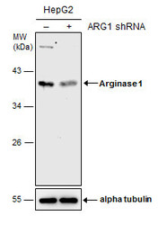
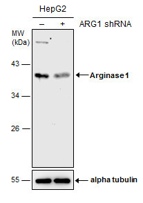
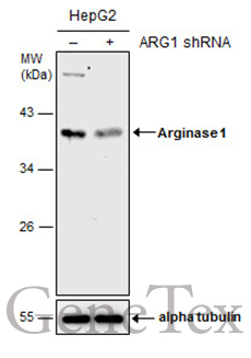
- Non-transfected (¡V) and transfected (+) HepG2 whole cell extracts (30 ?g) were separated by 10% SDS-PAGE, and the membrane was blotted with Arginase 1 antibody (GTX109242) diluted at 1:1000. The HRP-conjugated anti-rabbit IgG antibody (GTX213110-01) was used to detect the primary antibody.
Supportive validation
- Submitted by
- GeneTex (provider)
- Main image
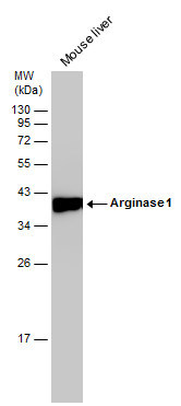
- Experimental details
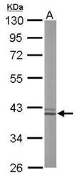
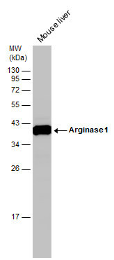
- Sample (20 ug of whole cell lysate) A: mouse liver 10% SDS PAGE GTX109242 diluted at 1:50000
- Validation comment
- WB

- Submitted by
- GeneTex (provider)
- Main image

- Experimental details
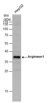
- Arginase 1 antibody detects ARG1 protein by Western blot analysis.A. 30 µg HepG2 whole cell lysate/extract10 % SDS-PAGEArginase 1 antibody (GTX109242) dilution: 1:1000
- Validation comment
- WB

- Submitted by
- GeneTex (provider)
- Main image

- Experimental details
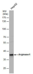
- Arginase 1 antibody detects Arginase 1 protein by western blot analysis. Whole cell extracts (30 ?g) was separated by 12% SDS-PAGE, and the membrane was blotted with Arginase 1 antibody (GTX109242) at a dilution of 1:1000.
- Validation comment
- WB
- Submitted by
- GeneTex (provider)
- Main image
- Experimental details
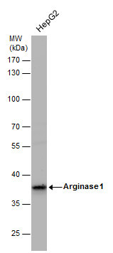
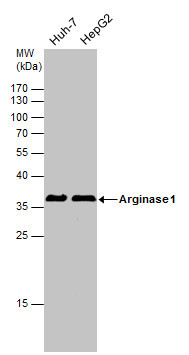
- Arginase 1 antibody detects Arginase 1 protein by western blot analysis. Whole cell extracts (30 ?g) was separated by 10% SDS-PAGE, and the membrane was blotted with Arginase 1 antibody (GTX109242) at a dilution of 1:1000.
- Validation comment
- WB
- Submitted by
- GeneTex (provider)
- Main image
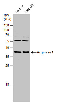
- Experimental details
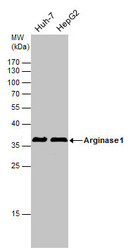
- Arginase 1 antibody detects Arginase 1 protein by western blot analysis. Various whole cell extracts (30 ?g) were separated by 12% SDS-PAGE, and the membrane was blotted with Arginase 1 antibody (GTX109242) diluted at 1:1000. The HRP-conjugated anti-rabbit IgG antibody (GTX213110-01) was used to detect the primary antibody.
- Submitted by
- GeneTex (provider)
- Main image

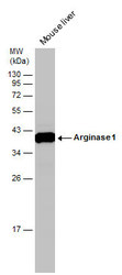
- Experimental details
- Mouse tissue extract (50 ?g) was separated by 12% SDS-PAGE, and the membrane was blotted with Arginase 1 antibody (GTX109242) diluted at 1:10000. The HRP-conjugated anti-rabbit IgG antibody (GTX213110-01) was used to detect the primary antibody.
- Submitted by
- GeneTex (provider)
- Main image

- Experimental details
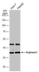
- Various whole cell extracts (30 £gg) were separated by 12% SDS-PAGE, and the membrane was blotted with Arginase 1 antibody (GTX109242) diluted at1:1000.
- Submitted by
- GeneTex (provider)
- Main image
- Experimental details
- Mouse tissue extract (50 ?g) was separated by 12% SDS-PAGE, and the membrane was blotted with Arginase 1 antibody (GTX109242) diluted at 1:10000.
- Submitted by
- GeneTex (provider)
- Main image

- Experimental details
- Non-transfected (¡V) and transfected (+) HepG2 whole cell extracts (30 ?g) were separated by 10% SDS-PAGE, and the membrane was blotted with Arginase 1 antibody (GTX109242) diluted at 1:1000. The HRP-conjugated anti-rabbit IgG antibody (GTX213110-01) was used to detect the primary antibody.
- Submitted by
- GeneTex (provider)
- Main image
- Experimental details
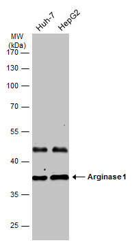
- Various whole cell extracts (30 ?g) were separated by 10% SDS-PAGE, and the membrane was blotted with Arginase 1 antibody (GTX109242) diluted at 1:1000. The HRP-conjugated anti-rabbit IgG antibody (GTX213110-01) was used to detect the primary antibody.
Supportive validation
- Submitted by
- GeneTex (provider)
- Main image

- Experimental details
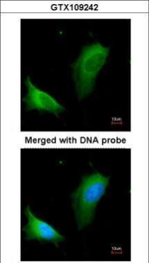
- Immunofluorescence analysis of paraformaldehyde-fixed HeLa, using arginase I (GTX109242) antibody at 1:200 dilution.
Supportive validation
- Submitted by
- GeneTex (provider)
- Main image
- Experimental details
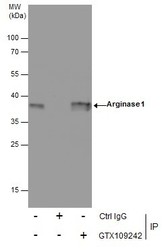
- Immunoprecipitation of Arginase 1 protein from HepG2 whole cell extracts using 5 ?g of Arginase 1 antibody (GTX109242).Western blot analysis was performed using Arginase 1 antibody (GTX109242).EasyBlot anti-Rabbit IgG (GTX221666-01) was used as a secondary reagent.
- Validation comment
- IP
- Submitted by
- GeneTex (provider)
- Main image

- Experimental details
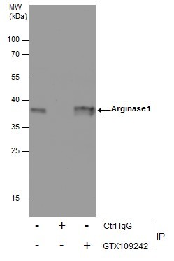
- Immunoprecipitation of Arginase 1 protein from HepG2 whole cell extracts using 5 £gg of Arginase 1 antibody (GTX109242).Western blot analysis was performed using Arginase 1 antibody (GTX109242).EasyBlot anti-Rabbit IgG (GTX221666-01) was used as a secondary reagent.
Supportive validation
- Submitted by
- GeneTex (provider)
- Main image
- Experimental details
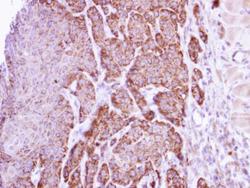
- Immunohistochemical analysis of paraffin-embedded Cal27 xenograft, using arginase I (GTX109242) antibody at 1:500 dilution.

- Submitted by
- GeneTex (provider)
- Main image
- Experimental details
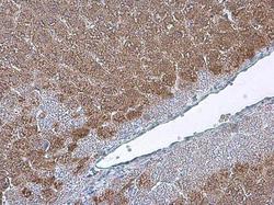
- Arginase 1 antibody detects Arginase 1 protein at cytoplasm in mouse liver by immunohistochemical analysis. Sample: Paraffin-embedded mouse liver. Arginase 1 antibody (GTX109242) diluted at 1:2500.

Supportive validation
- Submitted by
- GeneTex (provider)
- Main image

- Experimental details
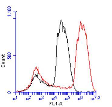
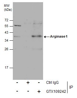
- Arginase 1 antibody (GTX109242) detects ARG1 protein by flow cytometry analysis. Sample: HepG2 cell. Black: Unlabelled sample was used as a control. Red: Arginase 1 antibody (GTX109242) dilution: 1:50. Acquisition of 20,000 events were collected using a Dylight 488-conjugated secondary antibody for FACS analysis.