 Explore
 Validate
 Learn
Learn Western blot
Antibody data
- Antibody Data
- Antigen structure
- References [6]
- Comments [0]
- Validations
- Western blot [3]
- Immunohistochemistry [5]
Submit
Validation data
Reference
Comment
Report error
- Product number
- NBP1-87490 - Provider product page

- Provider
- Novus Biologicals
- Proper citation
- Novus Cat#NBP1-87490, RRID:AB_11029644
- Product name
- Rabbit Polyclonal Arginase 1/ARG1/liver Arginase Antibody
- Antibody type
- Polyclonal
- Description
- Immunogen affinity purified. Specificity of human Arginase 1/ARG1/liver Arginase antibody verified on a Protein Array containing target protein plus 383 other non-specific proteins.
- Reactivity
- Human
- Host
- Rabbit
- Isotype
- IgG
- Vial size
- 0.1 ml
- Storage
- Store at 4C short term. Aliquot and store at -20C long term. Avoid freeze-thaw cycles.
Submitted references Arginase regulates red blood cell nitric oxide synthase and export of cardioprotective nitric oxide bioactivity.
Local arginase inhibition during early reperfusion mediates cardioprotection via increased nitric oxide production.
Arginase inhibition improves endothelial function in patients with coronary artery disease and type 2 diabetes mellitus.
Arginase inhibition restores in vivo coronary microvascular function in type 2 diabetic rats.
Arginase-1: a new immunohistochemical marker of hepatocytes and hepatocellular neoplasms.
A quantitative proteomic approach for identification of potential biomarkers in hepatocellular carcinoma.
Yang J, Gonon AT, Sjöquist PO, Lundberg JO, Pernow J
Proceedings of the National Academy of Sciences of the United States of America 2013 Sep 10;110(37):15049-54
Proceedings of the National Academy of Sciences of the United States of America 2013 Sep 10;110(37):15049-54
Local arginase inhibition during early reperfusion mediates cardioprotection via increased nitric oxide production.
Gonon AT, Jung C, Katz A, Westerblad H, Shemyakin A, Sjöquist PO, Lundberg JO, Pernow J
PloS one 2012;7(7):e42038
PloS one 2012;7(7):e42038
Arginase inhibition improves endothelial function in patients with coronary artery disease and type 2 diabetes mellitus.
Shemyakin A, Kövamees O, Rafnsson A, Böhm F, Svenarud P, Settergren M, Jung C, Pernow J
Circulation 2012 Dec 18;126(25):2943-50
Circulation 2012 Dec 18;126(25):2943-50
Arginase inhibition restores in vivo coronary microvascular function in type 2 diabetic rats.
Grönros J, Jung C, Lundberg JO, Cerrato R, Ostenson CG, Pernow J
American journal of physiology. Heart and circulatory physiology 2011 Apr;300(4):H1174-81
American journal of physiology. Heart and circulatory physiology 2011 Apr;300(4):H1174-81
Arginase-1: a new immunohistochemical marker of hepatocytes and hepatocellular neoplasms.
Yan BC, Gong C, Song J, Krausz T, Tretiakova M, Hyjek E, Al-Ahmadie H, Alves V, Xiao SY, Anders RA, Hart JA
The American journal of surgical pathology 2010 Aug;34(8):1147-54
The American journal of surgical pathology 2010 Aug;34(8):1147-54
A quantitative proteomic approach for identification of potential biomarkers in hepatocellular carcinoma.
Chaerkady R, Harsha HC, Nalli A, Gucek M, Vivekanandan P, Akhtar J, Cole RN, Simmers J, Schulick RD, Singh S, Torbenson M, Pandey A, Thuluvath PJ
Journal of proteome research 2008 Oct;7(10):4289-98
Journal of proteome research 2008 Oct;7(10):4289-98
No comments: Submit comment
Supportive validation
- Submitted by
- Novus Biologicals (provider)
- Main image

- Experimental details
- Simple Western: Arginase 1/ARG1/liver Arginase Antibody [NBP1-87490] - Simple Western lane view shows a specific band for ARG1 in 0.2 mg/ml of Liver (left), HepG2 (right) lysate. This experiment was performed under reducing conditions using the 12-230 kDa separation system.

- Submitted by
- Novus Biologicals (provider)
- Main image
- Experimental details
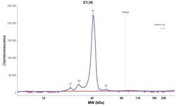
- Simple Western: Arginase 1/ARG1/liver Arginase Antibody [NBP1-87490] - Electropherogram image(s) of corresponding Simple Western lane view. Arginase 1/ARG1/liver Arginase antibody was used at 1:50 dilution on Liver and HepG2 lysate(s).

- Submitted by
- Novus Biologicals (provider)
- Main image

- Experimental details
- Western Blot: Arginase 1/ARG1/liver Arginase Antibody [NBP1-87490] - Analysis using Anti-ARG1 antibody NBP1-87490 (A) shows similar pattern to independent antibody NBP1-87455 (B).

Supportive validation
- Submitted by
- Novus Biologicals (provider)
- Main image

- Experimental details
- Immunohistochemistry-Paraffin: Arginase 1/ARG1/liver Arginase Antibody [NBP1-87490] - Staining of human bone marrow shows strong cytoplasmic and nuclear positivity in a subset of hematopoietic cells.

- Submitted by
- Novus Biologicals (provider)
- Main image

- Experimental details
- Immunohistochemistry-Paraffin: Arginase 1/ARG1/liver Arginase Antibody [NBP1-87490] - Staining of human hepatocellular carcinoma shows moderate cytoplasmic and nuclear positivity in tumor cells.

- Submitted by
- Novus Biologicals (provider)
- Main image

- Experimental details
- Immunohistochemistry-Paraffin: Arginase 1/ARG1/liver Arginase Antibody [NBP1-87490] - Staining of human liver shows strong cytoplasmic and nuclear positivity in hepatocytes.

- Submitted by
- Novus Biologicals (provider)
- Main image

- Experimental details
- Immunohistochemistry-Paraffin: Arginase 1/ARG1/liver Arginase Antibody [NBP1-87490] - Staining of human kidney shows no positivity in cells in tubules as expected.

- Submitted by
- Novus Biologicals (provider)
- Main image

- Experimental details
- Immunohistochemistry-Paraffin: Arginase 1/ARG1/liver Arginase Antibody [NBP1-87490] - Staining in human liver and kidney tissues . Corresponding ARG1 RNA-seq data are presented for the same tissues.
