 Explore
 Validate
 Learn
Learn Western blot
Antibody data
- Antibody Data
- Antigen structure
- References [6]
- Comments [0]
- Validations
- Western blot [3]
- Immunocytochemistry [2]
Submit
Validation data
Reference
Comment
Report error
- Product number
- 701161 - Provider product page

- Provider
- Invitrogen Antibodies
- Product name
- Occludin Recombinant Rabbit Monoclonal Antibody (6H10L9)
- Antibody type
- Monoclonal
- Antigen
- Synthetic peptide
- Reactivity
- Human
- Host
- Rabbit
- Isotype
- IgG
- Antibody clone number
- 6H10L9
- Vial size
- 100 µg
- Concentration
- 0.5 mg/mL
- Storage
- Store at 4°C short term. For long term storage, store at -20°C, avoiding freeze/thaw cycles.
Submitted references Early Activation of MAPK p44/42 Is Partially Involved in DON-Induced Disruption of the Intestinal Barrier Function and Tight Junction Network.
Kiwifruit cysteine protease actinidin compromises the intestinal barrier by disrupting tight junctions.
Heme oxygenase-1 protects corexit 9500A-induced respiratory epithelial injury across species.
Label-free recognition of drug resistance via impedimetric screening of breast cancer cells.
Claudin 1 expression characterizes human uterine cervical reserve cells.
Distinguishing drug-induced minor morphological changes from major cellular damage via label-free impedimetric toxicity screening.
Springler A, Hessenberger S, Schatzmayr G, Mayer E
Toxins 2016 Sep 8;8(9)
Toxins 2016 Sep 8;8(9)
Kiwifruit cysteine protease actinidin compromises the intestinal barrier by disrupting tight junctions.
Grozdanovic MM, Čavić M, Nešić A, Andjelković U, Akbari P, Smit JJ, Gavrović-Jankulović M
Biochimica et biophysica acta 2016 Mar;1860(3):516-26
Biochimica et biophysica acta 2016 Mar;1860(3):516-26
Heme oxygenase-1 protects corexit 9500A-induced respiratory epithelial injury across species.
Li FJ, Duggal RN, Oliva OM, Karki S, Surolia R, Wang Z, Watson RD, Thannickal VJ, Powell M, Watts S, Kulkarni T, Batra H, Bolisetty S, Agarwal A, Antony VB
PloS one 2015;10(4):e0122275
PloS one 2015;10(4):e0122275
Label-free recognition of drug resistance via impedimetric screening of breast cancer cells.
Eker B, Meissner R, Bertsch A, Mehta K, Renaud P
PloS one 2013;8(3):e57423
PloS one 2013;8(3):e57423
Claudin 1 expression characterizes human uterine cervical reserve cells.
Zinner B, Gyöngyösi B, Babarczi E, Kiss A, Sobel G
The journal of histochemistry and cytochemistry : official journal of the Histochemistry Society 2013 Dec;61(12):880-8
The journal of histochemistry and cytochemistry : official journal of the Histochemistry Society 2013 Dec;61(12):880-8
Distinguishing drug-induced minor morphological changes from major cellular damage via label-free impedimetric toxicity screening.
Meissner R, Eker B, Kasi H, Bertsch A, Renaud P
Lab on a chip 2011 Jul 21;11(14):2352-61
Lab on a chip 2011 Jul 21;11(14):2352-61
No comments: Submit comment
Supportive validation
- Submitted by
- Invitrogen Antibodies (provider)
- Main image

- Experimental details
- Western blot analysis of Occludin in HEK293 (lane 1) and MCF-7 (lane 2) whole cell extracts using an Occludin recombinant rabbit monoclonal antibody (Product # 701161) at a dilution of 2 µg/mL. Samples were detected using chemiluminescence (ECL). Results show a band at ~59kDa.

- Submitted by
- Invitrogen Antibodies (provider)
- Main image

- Experimental details
- Western blot analysis of Occludin was performed by loading 30 µg of HEK-293 and Hep G2 cell lysates using Novex®NuPAGE®4-12% Bis-Tris gel (Product # NP0321BOX), XCell SureLock Electrophoresis System (Product # EI0002), Novex® Sharp Pre-Stained Protein Standard (Product # LC5800), and iBlot® Dry Blotting System (Product # IB21001). Proteins were transferred to a nitrocellulose membrane and blocked with 5% skim milk for 1 hour at room temperature. Occludin was detected at ~59 kDa using Occludin Recombinant Rabbit Monoclonal Antibody (Product # 701161) at a 1:1000 dilution in 2.5% skim milk at 4°C overnight on a rocking platform. Detection was performed using an HRP-conjugated Goat anti-Rabbit secondary antibody (Product # G-21234) at a 1:5000 dilution and chemiluminescent detection was performed using Pierce™ ECL Western blotting Substrate (Product # 32106).

- Submitted by
- Invitrogen Antibodies (provider)
- Main image

- Experimental details
- Western blot analysis of Occludin in HEK293 (lane 1) and MCF-7 (lane 2) whole cell extracts using an Occludin recombinant rabbit monoclonal antibody (Product # 701161) at a dilution of 2 µg/mL. Samples were detected using chemiluminescence (ECL). Results show a band at ~59kDa.

Supportive validation
- Submitted by
- Invitrogen Antibodies (provider)
- Main image
- Experimental details
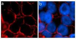
- Immunofluorescent analysis of Occludin in U2OS cells using an Occludin recombinant rabbit monoclonal antibody (Product # 701161) followed by detection using an Alexa Fluor 594 goat anti-rabbit secondary antibody (red) (Image A). Image B is a composite image with nuclei stained using DAPI (blue) showing localization of occludin at tight junctions.

- Submitted by
- Invitrogen Antibodies (provider)
- Main image

- Experimental details
- Immunofluorescent analysis of Occludin was performed on 90% confluent log phase Caco-2 cells. The cells were fixed with 4% paraformaldehyde for 15 minutes, and blocked with 5% BSA for 1 hour at room temperature. The cells were labeled with Occludin Recombinant Rabbit Monoclonal Antibody (Product # 701161) at a dilution of 1:500 in 1% BSA and incubated for 3 hours at room temperature and then labeled with Alexa Fluor® 488 Goat anti-Rabbit IgG secondary antibody (Product # A-11008) at a dilution of 1:400 for 30 minutes at room temperature (Panel a: green). Nuclei (Panel b: blue) were stained with SlowFade® Gold Antifade Mountant with DAPI (Product # S36938). Panel c is a merged image and showing cell junction localization and panel d is a control without primary antibody. The images were captured using a Nikon microscope at 20X magnification.
