 Explore
 Validate
 Learn
Learn Western blot
Antibody data
- Antibody Data
- Antigen structure
- References [3]
- Comments [0]
- Validations
- Western blot [1]
- Immunocytochemistry [1]
- Immunohistochemistry [1]
Submit
Validation data
Reference
Comment
Report error
- Product number
- AF4580 - Provider product page

- Provider
- R&D Systems
- Product name
- Mouse ESGP Antibody
- Antibody type
- Polyclonal
- Description
- Antigen Affinity-purified. Detects mouse ESGP in direct ELISAs and Western blots.
- Reactivity
- Mouse
- Host
- Sheep
- Conjugate
- Unconjugated
- Antigen sequence
Q2Q5T5- Isotype
- IgG
- Vial size
- 100 ug
- Concentration
- LYOPH
- Storage
- Use a manual defrost freezer and avoid repeated freeze-thaw cycles. 12 months from date of receipt, -20 to -70 °C as supplied. 1 month, 2 to 8 °C under sterile conditions after reconstitution. 6 months, -20 to -70 °C under sterile conditions after reconstitution.
Submitted references Fusogenic micropeptide Myomixer is essential for satellite cell fusion and muscle regeneration.
Myomerger induces fusion of non-fusogenic cells and is required for skeletal muscle development.
The microprotein Minion controls cell fusion and muscle formation.
Bi P, McAnally JR, Shelton JM, Sánchez-Ortiz E, Bassel-Duby R, Olson EN
Proceedings of the National Academy of Sciences of the United States of America 2018 Apr 10;115(15):3864-3869
Proceedings of the National Academy of Sciences of the United States of America 2018 Apr 10;115(15):3864-3869
Myomerger induces fusion of non-fusogenic cells and is required for skeletal muscle development.
Quinn ME, Goh Q, Kurosaka M, Gamage DG, Petrany MJ, Prasad V, Millay DP
Nature communications 2017 Jun 1;8:15665
Nature communications 2017 Jun 1;8:15665
The microprotein Minion controls cell fusion and muscle formation.
Zhang Q, Vashisht AA, O'Rourke J, Corbel SY, Moran R, Romero A, Miraglia L, Zhang J, Durrant E, Schmedt C, Sampath SC, Sampath SC
Nature communications 2017 Jun 1;8:15664
Nature communications 2017 Jun 1;8:15664
No comments: Submit comment
Supportive validation
- Submitted by
- R&D Systems (provider)
- Main image

- Experimental details
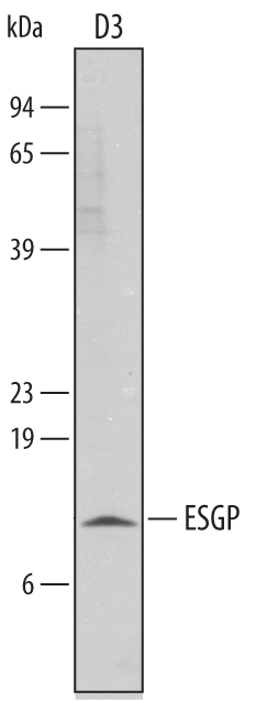
- Detection of Mouse ESGP by Western Blot. Western blot shows lysates of D3 mouse embryonic stem cell line. PVDF membrane was probed with 0.1 µg/mL of Sheep Anti-Mouse ESGP Antigen Affinity-purified Polyclonal Antibody (Catalog # AF4580) followed by HRP-conjugated Anti-Sheep IgG Secondary Antibody (Catalog # HAF016). A specific band was detected for ESGP at approximately 9 kDa (as indicated). This experiment was conducted under reducing conditions and using Immunoblot Buffer Group 8.
Supportive validation
- Submitted by
- R&D Systems (provider)
- Main image

- Experimental details
- ESGP in D3 Mouse Cell Line. ESGP was detected in immersion fixed D3 mouse embryonic stem cell line using Sheep Anti-Mouse ESGP Antigen Affinity-purified Polyclonal Antibody (Catalog # AF4580) at 10 µg/mL for 3 hours at room temperature. Cells were stained using the NorthernLights™ 557-conjugated Anti-Sheep IgG Secondary Antibody (red; Catalog # NL010) and counterstained with DAPI (blue). Specific staining was localized to cytoplasm and cell secretion. View our protocol for Fluorescent ICC Staining of Stem Cells on Coverslips.

Supportive validation
- Submitted by
- R&D Systems (provider)
- Main image
- Experimental details
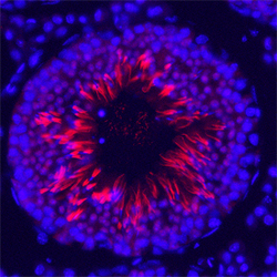
- ESGP in Mouse Testis. ESGP was detected in perfusion fixed frozen sections of mouse testis using Sheep Anti-Mouse ESGP Antigen Affinity-purified Polyclonal Antibody (Catalog # AF4580) at 1.7 µg/mL overnight at 4 °C. Tissue was stained using the Northern-Lights™ 557-conjugated Anti-Sheep IgG Secondary Antibody (red; Catalog # NL010) and counterstained with DAPI (blue). Specific staining was localized to late spermatids. View our protocol for Fluorescent IHC Staining of Frozen Tissue Sections.
