 Explore
 Validate
 Learn
Learn Immunohistochemistry
Antibody data
- Antibody Data
- Antigen structure
- References [2]
- Comments [0]
- Validations
- Immunohistochemistry [8]
Submit
Validation data
Reference
Comment
Report error
- Product number
- HPA035364 - Provider product page

- Provider
- Atlas Antibodies
- Proper citation
- Atlas Antibodies Cat#HPA035364, RRID:AB_10601095
- Product name
- Anti-CCDC39
- Antibody type
- Polyclonal
- Description
- Affinity purified using the PrEST antigen as affinity ligand
- Reactivity
- Human
- Host
- Rabbit
- Conjugate
- Unconjugated
- Antigen sequence
ILDNELTETISAQLELDKAAQDFRKIHNERQELIK
QWENTIEQMQKRDGDIDNCALELARIKQETREKEN
LVKEKIKFLESEIGNNTEFEKRISVADR- Isotype
- IgG
- Vial size
- 100 µl
- Storage
- Store at +4°C for short term storage. Long time storage is recommended at -20°C.
Submitted references DYX1C1 is required for axonemal dynein assembly and ciliary motility.
CCDC39 is required for assembly of inner dynein arms and the dynein regulatory complex and for normal ciliary motility in humans and dogs
Tarkar A, Loges NT, Slagle CE, Francis R, Dougherty GW, Tamayo JV, Shook B, Cantino M, Schwartz D, Jahnke C, Olbrich H, Werner C, Raidt J, Pennekamp P, Abouhamed M, Hjeij R, Köhler G, Griese M, Li Y, Lemke K, Klena N, Liu X, Gabriel G, Tobita K, Jaspers M, Morgan LC, Shapiro AJ, Letteboer SJ, Mans DA, Carson JL, Leigh MW, Wolf WE, Chen S, Lucas JS, Onoufriadis A, Plagnol V, Schmidts M, Boldt K, UK10K, Roepman R, Zariwala MA, Lo CW, Mitchison HM, Knowles MR, Burdine RD, Loturco JJ, Omran H
Nature genetics 2013 Sep;45(9):995-1003
Nature genetics 2013 Sep;45(9):995-1003
CCDC39 is required for assembly of inner dynein arms and the dynein regulatory complex and for normal ciliary motility in humans and dogs
Merveille A, Davis E, Becker-Heck A, Legendre M, Amirav I, Bataille G, Belmont J, Beydon N, Billen F, Clément A, Clercx C, Coste A, Crosbie R, de Blic J, Deleuze S, Duquesnoy P, Escalier D, Escudier E, Fliegauf M, Horvath J, Hill K, Jorissen M, Just J, Kispert A, Lathrop M, Loges N, Marthin J, Momozawa Y, Montantin G, Nielsen K, Olbrich H, Papon J, Rayet I, Roger G, Schmidts M, Tenreiro H, Towbin J, Zelenika D, Zentgraf H, Georges M, Lequarré A, Katsanis N, Omran H, Amselem S
Nature Genetics 2010 December;43(1):72-78
Nature Genetics 2010 December;43(1):72-78
No comments: Submit comment
Enhanced validation
Enhanced validation
Supportive validation
- Submitted by
- Atlas Antibodies (provider)
- Enhanced method
- Orthogonal validation
- Main image

- Experimental details
- Immunohistochemistry analysis in human testis and tonsil tissues using HPA035364 antibody. Corresponding CCDC39 RNA-seq data are presented for the same tissues.
- Sample type
- HUMAN

Enhanced validation
- Submitted by
- Atlas Antibodies (provider)
- Enhanced method
- Independent antibody validation
- Main image

- Experimental details
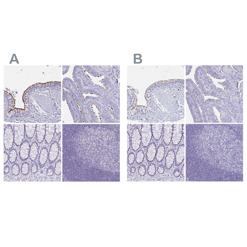
- Immunohistochemical staining of human bronchus, fallopian tube, rectum and tonsil using Anti-CCDC39 antibody HPA035364 (A) shows similar protein distribution across tissues to independent antibody HPA035363 (B).
Supportive validation
- Submitted by
- Atlas Antibodies (provider)
- Main image

- Experimental details
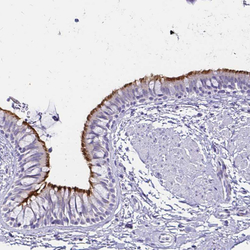
- Immunohistochemical staining of human bronchus shows strong positivity in cilia of respiratory epithelial cells.

- Submitted by
- Atlas Antibodies (provider)
- Main image
- Experimental details
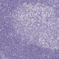
- Immunohistochemical staining of human tonsil shows no positivity in lymphoid cells as expected.
- Sample type
- HUMAN

- Submitted by
- Atlas Antibodies (provider)
- Main image

- Experimental details
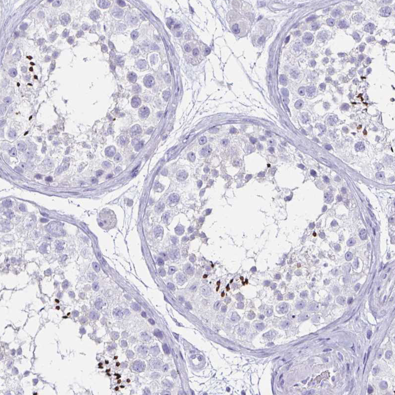
- Immunohistochemical staining of human testis shows strong cytoplasmic positivity in spermatozoa.
- Sample type
- HUMAN
- Submitted by
- Atlas Antibodies (provider)
- Main image
- Experimental details
- Immunohistochemical staining of human bronchus shows strong positivity in cilia in glandular cells.
- Sample type
- HUMAN

- Submitted by
- Atlas Antibodies (provider)
- Main image
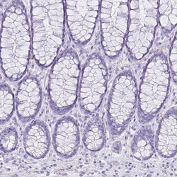
- Experimental details
- Immunohistochemical staining of human rectum shows no positivity in glandular cells as expected.
- Sample type
- HUMAN

- Submitted by
- Atlas Antibodies (provider)
- Main image

- Experimental details
- Immunohistochemical staining of human fallopian tube shows strong positivity in cilia in glandular cells.
- Sample type
- HUMAN
