Antibody data
- Antibody Data
- Antigen structure
- References [10]
- Comments [0]
- Validations
- Immunocytochemistry [1]
- Immunohistochemistry [1]
- Flow cytometry [1]
Submit
Validation data
Reference
Comment
Report error
- Product number
- MA5-11454 - Provider product page

- Provider
- Invitrogen Antibodies
- Product name
- Thrombomodulin Monoclonal Antibody (141C01 (1009))
- Antibody type
- Monoclonal
- Antigen
- Recombinant full-length protein
- Description
- MA5-11454 targets CD141 in IHC (P), FACS and ICC/IF applications and shows reactivity with Human and Rat samples. The MA5-11454 immunogen is recombinant protein encoding the six repeated EGF domains of human thrombomodulin.
- Reactivity
- Human, Rat
- Host
- Mouse
- Isotype
- IgG
- Antibody clone number
- 141C01 (1009)
- Vial size
- 500 µL
- Concentration
- Conc. Not Determined
- Storage
- 4° C
Submitted references Cellular and gene signatures of tumor-infiltrating dendritic cells and natural-killer cells predict prognosis of neuroblastoma.
Low grade urothelial carcinoma mimicking basal cell hyperplasia and transitional metaplasia in needle prostate biopsy.
The endothelial microenvironment in the venous valvular sinus: thromboresistance trends and inter-individual variation.
The urokinase receptor supports tumorigenesis of human malignant pleural mesothelioma cells.
Cellular regulation of blood coagulation: a model for venous stasis.
Valves of the deep venous system: an overlooked risk factor.
Role of impaired peritubular capillary and hypoxia in progressive interstitial fibrosis after 56 subtotal nephrectomy of rats.
Healthy and pre-eclamptic placental basal plate lining cells: quantitative comparisons based on confocal laser scanning microscopy.
Confocal laser scanning microscope study of cytokeratin immunofluorescence differences between villous and extravillous trophoblast: cytokeratin downregulation in pre-eclampsia.
Cell fusion-independent differentiation of neural stem cells to the endothelial lineage.
Melaiu O, Chierici M, Lucarini V, Jurman G, Conti LA, De Vito R, Boldrini R, Cifaldi L, Castellano A, Furlanello C, Barnaba V, Locatelli F, Fruci D
Nature communications 2020 Nov 25;11(1):5992
Nature communications 2020 Nov 25;11(1):5992
Low grade urothelial carcinoma mimicking basal cell hyperplasia and transitional metaplasia in needle prostate biopsy.
Arista-Nasr J, Martinez-Benitez B, Bornstein-Quevedo L, Aguilar-Ayala E, Aleman-Sanchez CN, Ortiz-Bautista R
International braz j urol : official journal of the Brazilian Society of Urology 2016 Mar-Apr;42(2):247-52
International braz j urol : official journal of the Brazilian Society of Urology 2016 Mar-Apr;42(2):247-52
The endothelial microenvironment in the venous valvular sinus: thromboresistance trends and inter-individual variation.
Trotman WE, Taatjes DJ, Callas PW, Bovill EG
Histochemistry and cell biology 2011 Feb;135(2):141-52
Histochemistry and cell biology 2011 Feb;135(2):141-52
The urokinase receptor supports tumorigenesis of human malignant pleural mesothelioma cells.
Tucker TA, Dean C, Komissarov AA, Koenig K, Mazar AP, Pendurthi U, Allen T, Idell S
American journal of respiratory cell and molecular biology 2010 Jun;42(6):685-96
American journal of respiratory cell and molecular biology 2010 Jun;42(6):685-96
Cellular regulation of blood coagulation: a model for venous stasis.
Campbell JE, Brummel-Ziedins KE, Butenas S, Mann KG
Blood 2010 Dec 23;116(26):6082-91
Blood 2010 Dec 23;116(26):6082-91
Valves of the deep venous system: an overlooked risk factor.
Brooks EG, Trotman W, Wadsworth MP, Taatjes DJ, Evans MF, Ittleman FP, Callas PW, Esmon CT, Bovill EG
Blood 2009 Aug 6;114(6):1276-9
Blood 2009 Aug 6;114(6):1276-9
Role of impaired peritubular capillary and hypoxia in progressive interstitial fibrosis after 56 subtotal nephrectomy of rats.
Zhang B, Liang X, Shi W, Ye Z, He C, Hu X, Liu S
Nephrology (Carlton, Vic.) 2005 Aug;10(4):351-7
Nephrology (Carlton, Vic.) 2005 Aug;10(4):351-7
Healthy and pre-eclamptic placental basal plate lining cells: quantitative comparisons based on confocal laser scanning microscopy.
Smith RK, Ockleford CD, Byrne S, Bosio P, Sanders R
Microscopy research and technique 2004 May 1;64(1):54-62
Microscopy research and technique 2004 May 1;64(1):54-62
Confocal laser scanning microscope study of cytokeratin immunofluorescence differences between villous and extravillous trophoblast: cytokeratin downregulation in pre-eclampsia.
Ockleford CD, Smith RK, Byrne S, Sanders R, Bosio P
Microscopy research and technique 2004 May 1;64(1):43-53
Microscopy research and technique 2004 May 1;64(1):43-53
Cell fusion-independent differentiation of neural stem cells to the endothelial lineage.
Wurmser AE, Nakashima K, Summers RG, Toni N, D'Amour KA, Lie DC, Gage FH
Nature 2004 Jul 15;430(6997):350-6
Nature 2004 Jul 15;430(6997):350-6
No comments: Submit comment
Supportive validation
- Submitted by
- Invitrogen Antibodies (provider)
- Main image

- Experimental details
- Immunofluorescent analysis of CD141 (green) showing staining in the membrane of A431 cells (right) compared to a negative control without primary antibody (left). Formalin-fixed cells were permeabilized with 0.1% Triton X-100 in TBS for 5-10 minutes and blocked with 3% BSA-PBS for 30 minutes at room temperature. Cells were probed with a CD141 monoclonal antibody (Product # MA5-11454) in 3% BSA-PBS at a dilution of 1:100 and incubated overnight at 4 ºC in a humidified chamber. Cells were washed with PBST and incubated with a DyLight-conjugated secondary antibody in PBS at room temperature in the dark. F-actin (red) was stained with a fluorescent red phalloidin and nuclei (blue) were stained with Hoechst or DAPI. Images were taken at a magnification of 60x.
Supportive validation
- Submitted by
- Invitrogen Antibodies (provider)
- Main image

- Experimental details
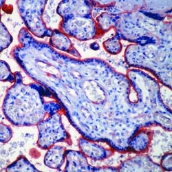
- Formalin-fixed, paraffin-embedded human placenta stained with CD141 antibody using peroxidase-conjugate and AEC chromogen. Note membrane staining of trophoblasts and endothelial cells.
Supportive validation
- Submitted by
- Invitrogen Antibodies (provider)
- Main image

- Experimental details
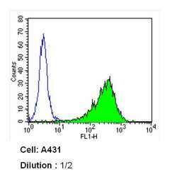
- Flow cytometry analysis of CD141 in A431 cells (green) compared to an isotype control (blue). Cells were harvested, adjusted to a concentration of 1-5x10^6 cells/mL, fixed with 2% paraformaldehyde and washed with PBS. Cells were blocked with a 2% solution of BSA-PBS for 30 min at room temperature and incubated with a CD141 monoclonal antibody (Product # MA5-11454) at a dilution of 1:2 for 60 min at room temperature. Cells were then incubated for 40 min at room temperature in the dark using a Dylight 488-conjugated secondary antibody and re-suspended in PBS for FACS analysis.
 Explore
 Validate
 Learn
Learn Immunocytochemistry