Antibody data
- Antibody Data
- Antigen structure
- References [0]
- Comments [0]
- Validations
- Western blot [1]
- Immunohistochemistry [1]
- Other assay [7]
Submit
Validation data
Reference
Comment
Report error
- Product number
- 32-6200 - Provider product page

- Provider
- Invitrogen Antibodies
- Product name
- Desmocollin 2/3 Monoclonal Antibody (7G6)
- Antibody type
- Monoclonal
- Antigen
- Other
- Reactivity
- Human
- Host
- Mouse
- Isotype
- IgG
- Antibody clone number
- 7G6
- Vial size
- 100 µg
- Concentration
- 0.5 mg/mL
- Storage
- -20°C
No comments: Submit comment
Supportive validation
- Submitted by
- Invitrogen Antibodies (provider)
- Main image

- Experimental details
- Western blot analysis of Desmocollin-2+3 was performed by loading 20 µg of PC-3 (lane1), COLO 205 (lane2), HeLa (lane3) and A431 (lane4) lysate using Novex®NuPAGE® 12 % Bis-Tris gel (Product # NP0342BOX), XCell SureLock Electrophoresis System (Product # EI0002), Novex® Sharp Pre-Stained Protein Standard (LC5800), and proteins were transferred to a PVDF membrane and blocked with 5% skim milk at 4°C overnight. Desmocollin-2+3 was detected at ~ 120, 105, 99 and 93 kDa using Desmocollin-2+3 Mouse Monoclonal Antibody (Product # 32-6200) at 1-2 µg/mL in 5% skim milk for 3 hour at room temperature on a rocking platform. Goat Anti-Mouse - HRP Secondary Antibody (Product # 62-6520) at 1:4000 dilution was used and chemiluminescent detection was performed using Pierce™ ECL Western Blotting Substrate (Product # 32106).
Supportive validation
- Submitted by
- Invitrogen Antibodies (provider)
- Main image

- Experimental details
- Immunohistochemistry analysis of Desmocollin-2/3 showing staining in the membrane of paraffin-embedded human heart tissue (right) compared to a negative control without primary antibody (left). To expose target proteins, antigen retrieval was performed using 10mM sodium citrate (pH 6.0), microwaved for 8-15 min. Following antigen retrieval, tissues were blocked in 3% H2O2-methanol for 15 min at room temperature, washed with ddH2O and PBS, and then probed with a Desmocollin-2/3 monoclonal antibody (Product # 32-6200) diluted in 3% BSA-PBS at a dilution of 1:20 overnight at 4ºC in a humidified chamber. Tissues were washed extensively in PBST and detection was performed using an HRP-conjugated secondary antibody followed by colorimetric detection using a DAB kit. Tissues were counterstained with hematoxylin and dehydrated with ethanol and xylene to prep for mounting.
Supportive validation
- Submitted by
- Invitrogen Antibodies (provider)
- Main image

- Experimental details
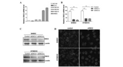
- Figure 1 DSC2 silencing in ESCC cells by siRNAs. (A) RT-qPCR analysis of the expression of DSC2 in ESCC cell lines. The expression levels of DSC2 were normalized to that of beta-actin. (B) RT-qPCR analysis of DSC2 silencing by siRNAs. Cells were transfected with DSC2 siRNA or control siRNA. (C) DSC2 silencing in SHEEC and KYSE510 cells was evaluated using western blot analysis. beta-actin served as a loading control. (D) Immunofluorescence analysis of DSC2 silencing by siRNAs (magnification, x400). DSC2, desmocollin-2; ESCC, esophageal squamous cell carcinoma; RT-qPCR, reverse transcription quantitative polymerase chain reaction.
- Submitted by
- Invitrogen Antibodies (provider)
- Main image

- Experimental details
- Figure 3 RNA interference-mediated inhibition of DSC2 affects desmosome protein expression and localization. (A) Immunofluorescence analysis of the subcellular localizations of the DSG2 and PKP2 proteins (magnification, x400). Of note, knocking down the expression of DSC2 caused reduced DSG2 and PKP2 membrane localization (white arrows). (B) Western blot analyses of DSG2 and PKP2 protein expression in the DSC2-specific siRNA or control-transfected SHEEC cells. beta-actin served as a loading control. DSC2, desmocollin-2; siDSC2, DSC2-specific siRNA; DSG2, desmoglein-2; PKP2, plakophilin-2.
- Submitted by
- Invitrogen Antibodies (provider)
- Main image

- Experimental details
- Figure 5 DSC2 depletion leads to keratin intermediate filament retraction and F-actin cytoskeleton rearrangement. (A) Immunofluorescence analysis shows retracted keratin intermediate filaments from plasma membranes in siDSC2-transfected SHEEC cells, whereas the plasma membranes of neighboring cells remained adjacent in the control-transfected SHEEC cells (magnification, x400). (B) SHEEC and KYSE510 cells were transiently transfected with control siRNA and siDSC2. The transfected cells were then fixed and F-actin organization was analyzed using phalloidin staining (magnification, x400). DSC2, desmocollin-2; siDSC2, DSC2-specific siRNA.
- Submitted by
- Invitrogen Antibodies (provider)
- Main image

- Experimental details
- Fig. 10. Studying the interactions between DSC2, PKP1, and VIM and their downstream molecules. ( A ) Western blot results revealing that treating A-SSP6 cells with the FAK inhibitor 14 (10 muM) or the Src inhibitor (50 nM) for 24 hours reduced the levels of FAK, p-Src, p-AKT, and p-ERK1/2. DMSO, dimethyl sulfoxide. ( B and C ) Western blotting of p-AKT, Bcl-2, p-ERK1/2, and ZEB1 in the A-SSP6 cells treated with the PI3K inhibitor dactolisib (1 muM) or MEK inhibitor trametinib (10 nM) for 24 hours. ( D ) Western blots showing the increases in VIM, ITGB1, FAK, p-Src, p-AKT, and p-ERK1/2 after overexpression of VIM in A-SSP6 cells with double knockdown of DSC2 and PKP1. ( E to G ) qRT-PCR results showing the knockdown effects of shDSC2 (E), shPKP1 (F), and shVIM (G) on the mRNA levels of DSC2, PKP1, VIM, and Bcl-2. As GAPDH is a house-keeping gene, it was used as an internal control for qRT-PCR experiments. The mRNA level in shCtrl group of A-SSP6 was normalized to 1. The results are the means +- SD from three independent experiments. Significant differences were determined by two-way ANOVA (E to G). * P < 0.05, ** P < 0.01, *** P < 0.001, and **** P < 0.0001.
- Submitted by
- Invitrogen Antibodies (provider)
- Main image

- Experimental details
- Fig. 4. Identification of adhesive molecules that were up-regulated in SSP6 cells. ( A ) Western blots showing the change in adhesive proteins in the M-SSP6 and A-SSP6 cells. Quantified data are presented as the means +- SD from three independent experiments. ( B ) Representative immunofluorescence (IF) staining images illustrating the distribution of DSC2, PKP1, vimentin (VIM), E-cadherin (CDH1), beta-catenin, fibronectin (FN1), and integrin beta 1 (ITGB1) in the A549-C3 and MCF7-C3 cells before and after SS15 treatment for 12 hours. Scale bar, 10 mum. ( C ) qRT-PCR results showing the normalized mRNA levels of DSC2, PKP1, VIM, CDH1, FN1, and ZEB1 in the A549-C3 cells under SS15 treatment at various time points. As glyceraldehyde-3-phosphate dehydrogenase (GAPDH) is a housekeeping gene whose mRNA level in A549-C3 cells at 0 hours was normalized to 1 and used as an internal reference for calculating the mRNA levels of the other tested genes. The results represent the means +- SD from three independent experiments. Significant differences were determined by one-way analysis of variance (ANOVA) (C). * P < 0.05, ** P < 0.01, *** P < 0.001, and **** P < 0.0001.
- Submitted by
- Invitrogen Antibodies (provider)
- Main image

- Experimental details
- Fig. 9. Signaling pathways of DSC2 and PKP1. ( A ) Western blots showing the higher expression of p-AKT, Bcl-2, and p-ERK1/2 in A-SSP6 cells. ( B ) Western blots showing the knockdown effects of DSC2, PKP1, DSC2 + PKP1, VIM, and ZEB1 on themselves and other proteins in A-SSP6 cells. ( C ) Western blots showing the effects of knockout ITGB1 on various indicated proteins in A-SSP6 cells. ( D ) DSC2 was overexpressed in A549-C3 cells and Western blots showing the up-regulation of VIM and ITGB1. ( E ) DSC2 was first overexpressed in A549-C3 cells, and then VIM was knocked down in these overexpressed cells, Western blots showing the down-regulation of VIM and ITGB1 after knockdown of VIM. ( F ) Western blot results indicating that shFN1 reduced the levels of ITGB1, FAK, and p-Src. ( G ) Western blotting showing that the levels of FAK and p-Src were decreased after VIM was knocked down. ( H ) The left side of the schematic illustration shows that CTCs with high levels of DSC2 can form clusters, survive in circulation, and subsequently form lung colonies by tail vein injection or metastasize to the liver, intestine, and brain in the lung orthotopic model. The right side of the schematics illustrates the proposed signaling pathways through which DSC2/PKP1 and VIM support cluster formation, cell survival in circulation, lung colony formation, and metastasis.
- Submitted by
- Invitrogen Antibodies (provider)
- Main image

- Experimental details
- FIGURE 2: Dsc2 is decreased in human and mouse inflamed colon, and its loss resulted in delayed mucosal recovery from colitis. (A) Representative images of IF staining of Dsc2 (green) and DAPI (blue) in frozen sections of human colonic tissues from patients with UC and healthy controls. Reduction of fluorescence intensity for Dsc2 in UC compared with control. Scale bars: 50 mum. (B) Western blot (WB) and densitometry analysis of the expression of Dsc2 and loading control Cytokeratin-8 (CK-8) in human colonic tissues from patients with UC and healthy controls. WB images are representative of two individual experiments with 10 controls and nine UC samples. Arrowhead: Full-length Dsc2; asterisk: cleaved Dsc2. Bar graphs represent densitometric values of seven healthy controls and six UC samples and are normalized to control. Data are mean +- SEM. Significance is determined by two-tailed Student's t test. * p
 Explore
 Validate
 Learn
Learn Western blot
 Immunoprecipitation